Chapter 6.5 Methods of stimulation and measurement 
|
CRANIAL ELECTROSTIMULATION (CES) |   | ||||
| THE MEANS WHEREBY | |||||
MA or EA? |    |
Focus on the needle | |||
To TENS or not to TENS? | 
| |||
| Studies where EA is more effective than TENS | | ||||
| Studies where EA and TENS are equally effective | ||||
| Studies where TENS is more effective than EA | ||||
| Studies where MA is more effective than TENS |
| ||||
| Studies where TENS is more effective than MA | ||||
| Other comparisons |
| ||||
| Other forms of electrical point or meridian stimulation | ||||
OTHER METHODS OF STIMULATING ACUPOINTS AND MERIDIANS |  | |||
|
Heat and cold Microwaves Millimetre waves (Extremely high frequency, EHF) Light | ||||||
| Low-intensity laser and polarised light |  
| ||||||
| Comparative studies of ‘laser acupuncture’ | ||||||
| Low-intensity pulsed electromagnetic fields |
| ||||||
|
Permanent magnets Vibration Sound Ultrasound | ||||
POSSIBLE ADVERSE EFFECTS OF STIMULATION | 
| |||
|
Electrical stimulation | ||||
| LILT |
| |||
| The vexed question of epilepsy |
| |||
Factors having possible adverse effects on stimulation |
| |||
|
The state of the patient Drug interactions Other treatments and environmental factors | |||
| EXPERIMENTAL MEASUREMENTS | |||
|
Electrodermal measurements at AcPs and TrPs in response to treatment: in search of the objective | |||
|
Preamble Back to the point Electroacupuncture according to Voll (EAV) | |||
|
Dutch EAV studies A British EAV study An American EAV study | |||
|
Other electrodermal measurement systems | |||
|
Measuring temperature Electrical imaging methods | |||
| Section summary | |||
|
Measurements of magnetic fields Box 6.5.1 A note on muscle and magnets | |||
In Conclusion | |||
| Other icons used in this chapter: |
 | |
| Cranial electrostimulation (CES) | | ||||
|
Auricular EA, so often used in the treatment of drug withdrawal, is not without precedent. As outlined in Ch. 2, the search for an effective electrical method for inducing analgesia, anaesthesia or generalised relaxation (‘electrosleep’) has resulted in a long tradition of applying stimulation to the head, generally via surface electrodes, although the relationships between the various methods used are not always clear.1 In the mid nineteenth century, Julius Althaus (see Ch. 4, ‘late-nineteenth-century clinicians’) applied DC between the forehead and occiput. Stéphane Leduc around 1900 first used interrupted DC applied on the midline between forehead and lumbar region, and then AC.2 Others, like a number of late nineteenth century clinicians,3 have explored linking forehead and leg4 (see Ch. 4, ‘late-nineteenth-century clinicians’) or forehead and wrist,5 and even mouth and anus6,7 (not recommended!). By the early 1950s, at least in the former Soviet Union, electrode positions had become standardised: two electrodes (generally cathodes) over the forehead or closed eyes, and two (the anodes) at the occiput. However, as one influential American researcher, Robert Smith, pointed out,8 in dogs it was a lot easier to get the electrodes to stay on if they were positioned bitemporally. With the development of auricular acupuncture, a transverse location became more popular, placed just in front of the top of the ears (Saul Liss), on the occiput just behind the ears (Margaret Patterson, Bob Beck), or of course between AcPs in the ears themselves (Wen Hsiang-lai, Lorenz Ng). In the former USSR the forehead/occiput arrangement remains common. One commercially available Swiss device even uses an intraoral electrode! Cranial and body AcP locations have been compared; Liss determined that changes in circulating neurochemicals were greater using the transcranial locations than LI-4 bilaterally, for instance.9 All in all, apart from the obvious division into two ‘families’ of CES, with electrodes positioned either ‘fore and aft’ or transversely, it would seem that the actual locations used for stimulation (whether AcPs or not) might be less significant than the electrical parameters of the stimulation,10 provided the factor of skin impedance is overcome.11 Perhaps because Leduc began by using 100 Hz, most Russian electrosleep (‘electronarcosis’ or ‘central electroanalgesia’) research also used this frequency, although probably only because, with the large currents involved at that time, it was high enough to avoid muscle twitching.12 Wen too used frequencies around 120–125 Hz (see SubCh. 6.3, ‘no effect on plasma or CSF βEP in addicts’), again only because of historical accident: the output of the first Chinese EA device that came to hand went no higher than 111 Hz.13 Even so, results of a number of early controlled studies seemed to favour 100 Hz14 over other frequencies, as has research on sinewave cranial TENS that supports the use of 100 Hz (as opposed to 5 Hz or 2 kHz), in terms of its effect both on blood pressure (BP) and heart rate (HR)15 and on experimental trigeminal pain.16 However, some researchers have felt that a slower, rhythmically repetitive stimulation would be more effective17,18 (see SubCh. 6.3, ‘animal hypnotism’), with frequencies around 100 Hz considered to be more ‘activating’.19 There are different interpretations of ‘low’ frequency in this context; in Russia, for instance, frequencies from 1–2 Hz up to 100–130 Hz are considered by some authorities as suited to inhibition of CNS activity,20 whereas in one small informal American study, 100 Hz (0.5–1.5 mA, 1 ms) was unexpectedly found to enhance alertness and even induce a sense of euphoria in some subjects.21 Applying strong currents transcranially can result in adverse effects such as spasm and even convulsion (below, ‘may well induce convulsions’). Attempts were made to reduce these by ramping current intensity,22 combining biphasic and monophasic currents (Anan’ev,23 Lebedev24,25 and others26), or using high (Knutson 700–1500 Hz) or very high frequencies (Liss 15 kHz, Kuster 20–100 MHz,27 Limoge 100 or 166 kHz), generally modulated at lower frequencies. For this purpose, Saul Liss has used 15 Hz (‘the bioactive frequency’28) and Aimé Limoge 100 Hz29 or 77 Hz30 (77 Hz has also been used in a Russian device31). Magnes’s Israeli group, for instance, found a 3.5–4.5 kHz sinewave modulated at 100 Hz gave analgesia together with signs of anaesthesia (stimulation through electrodes placed at various locations actually in the brain stem resulted in analgesia only).32 Liss, in experiments on GABA synthesis, defined the optimum parameters for his ‘Liss cranial stimulator’ as 15 kHz modulated at 15 Hz, adding in 500 Hz to reduce the amount of charge transferred by 50%33 (even though effects were not quite as good, changes in circulating neurochemical levels were better than those obtained using 80 Hz TENS34). Lebedev’s Moscow group, in contrast, found simple 70 Hz (3–3.5 ms) rectangular pulses combined with DC (with a ratio of DC to pulse current of 2:1) to be optimal for analgesia in rats as well as other animals.35 Some American authors considered 100 Hz sinewaves produce greater analgesia, with 700 Hz – 10 kHz resulting in deeper relaxation.36 Over the years, more and more CES devices have become available that have effects even when the currents applied are below the threshold for sensory stimulation (Ifor Capel, Daniel Kirsch), at 1 mA or below.37 Although no longer producing sufficient analgesia for surgical procedures (as with EAA, this requires strong stimulation), they have useful applications and are safe to use in everyday clinical practice as well as by patients at home. Even where electroanaesthesia is concerned, as one prominent early Russian researcher stated, ‘it is not the amount of electric current flowing through the brain that matters, but the excitatory, stimulating effect of a steep increase in current’.38 Thus waveform may in some circumstances be more important than intensity. Most CES systems use squarewaves in some form.39 However, one American device using 100 Hz sinewave stimulation has been well researched,40,41,42 a few have used random noise rather than repetitive signals43,44 and one a beat pattern between two different biphasic spike frequencies (0.4 and 0.5 Hz) that only repeats every 20 seconds or so.45 EEG-modulated trains have also been employed, giving better effects in cats (in terms of relaxation) than unmodulated trains.46 Treatment duration may also be an important factor: 20 minutes of Russian style CES inhibits high-threshold nociceptors, short (5-minute) or very short (1-minute) treatments inhibit only medium- or low-threshold mechanoreceptors respectively.47 Furthermore, in transverse CES systems using DC or asymmetric biphasic stimulation (even if charge balanced), which side of the head is positive (generally the right) can be an important factor.48,49 Indeed, it is noteworthy that unilateral stimulation may be quite ineffective using some forms of CES.50 Hence, whereas forehead to limb electrode arrangements may produce results (above, ‘forehead and wrist’), single ear to foot or tail may not.51 Ifor Capel, with colleagues in both Britain and the USA, has carried out a meticulous series of studies over more than 20 years using earlobe CES. He calls this SPES (for ‘subperception electrical stimulation’). Unlike many forms of CES, stimulation is applied via needle electrodes, but these only penetrate the epidermis (not the dermis52) sufficiently to overcome skin impedance differences. The stimulation itself is very precisely defined, and is one that Capel’s team has found to be very effective for pain. It consists of a biphasic rectangular charge-balanced waveform with a first (positive) phase duration of 2–3 ms, amplitude 10 µA and repetition rate 10 Hz. These parameters were arrived at after testing a wide range of options: frequencies of 7.5 or 50 Hz were virtually ineffective, for instance, and other currents in the 5–20 µA range also less effective,53,54 as were sine or spike waveforms.55,56 To test for pain, Capel’s coworkers have used the rat tail flick latency (TFL) model, among others (subthreshold vagal stimulation is well known to decrease TFL57). In contrast to Ng’s auricular EA (see SubCh. 6.3, ‘animal hypnotism’), the effects of this form of CES on opiate withdrawal treatment appear quite robust and immune to environmental disturbances. However, excessive noise or disturbance can affect results when working with pain, perhaps because of the sensitivity of the TFL model to stress (see SubCh. 6.3, ‘Stress also may invalidate HP or TFL testing’)58,59 (this is true of other pain measures as well, such as the radiant heat snout test60). SPES may indeed be more potent in its antinociceptive effects than focal electrical stimulation of the NRD or lH within the brain,61,62 with particular effects on nociceptive activity in the habenula,63 and NPf.64 SPES analgesia, like LF EA (see SubCh. 6.4, ‘a delayed onset’), is slow in onset (although this may not always be the case65) and with an after-effect that may last several hours, possibly with cycles of greater and less effect until dying away altogether.66 With SPES administered before anaesthetic agents such as hexobarbital, the 10 Hz signal also decreased the sleeping time of the anaesthetised rats, as did 500 Hz (although curiously only the latter was effective if applied peripherally via the paws).67 In contrast, pentobarbital may have a reverse effect, negating SPES antinociception if given before SPES is started.68,69 SPES may also affect blood sugar levels and appetite, in both rats and humans.70 The 10 Hz frequency (but with a stimulus very different from SPES in other respects) has also been found to enhance immune function in stressed rats, although 1 kHz proved more effective prophylactically.71 Other forms of CES may also have immune effects,72 with strong electronarcosis temporarily affecting lymphocyte: Neutrophil ratios, for example.73 A series of Japanese–American studies has explored the role of scalp sensory nerves in CES.74,75,76 Although the simplest pathway between cranial electrodes is over the skin surface, a certain amount of current (maybe a fifth,77 or even more78,79) does in fact penetrate the skull via more complex pathways (through the orbital and occipital foramina, via the auditory meatus, or by stimulating superficial branches of the cranial nerves – V, VII, IX or X in the case of auricular stimulation80). Thus even when electrodes are applied only on the forehead (in an attempt at ‘sham’ CES), current may penetrate the skull particularly if points of low skin resistance (SR) over cranial nerve branches are used.81 Skull thickness is not important.82 Also, if the cranial nerves are stimulated their cell bodies may be directly activated, with a cascade of effects in the brainstem where they are located; that some forms of unilateral CES (but not all, see above) are in fact effective would seem to support this possibility.83 The SPES signal in particular may penetrate the brain and directly affect the CNS,84 as preliminary peripheral nerve block studies did not seem to reduce its efficacy.85 This seems to be the case for other forms of CES as well. In rabbits, for instance (using 4 and 4.2 kHz with a resulting 200 Hz interference current), endogenous electrical activity was found to be affected primarily in the hypothalamus, thalamic centromedian nucleus (NCm), periaqueductal grey (PAG) matter and reticular formation – areas also implicated in EAA. However, in dead rabbits current only flowed over the skull surface.86 In rats, sinewave CES has been shown to affect metabolic activity in the PAG, solitary tract nucleus (NST), reticular formation, trigeminal nucleus and elsewhere.87 Theoretically, CES should affect the thalamus;88 indeed in monkeys some 42% of the applied current from strong (100 mA, 20–2000 Hz) TCET enters the brain as a whole, with slightly higher current concentrations measured in the thalamus compared with those in the cortex (although this difference may not be significant).89 Currents have also been detected during such treatment in the frontal lobes and brainstem.90 Methods have thus been developed for positioning electrodes in order to maximise current density,91,92 particularly in the thalamus.93 One study noted that synchronised 5–7 Hz discharges persisted in non-specific thalamic regions after 5–10 Hz CES, later spreading to other regions of the brain as well,94 and changes in thalamocortical EP with strong CES have also been investigated.95 Current changes have been measured too in the hippocampus and associated areas.96 With strong cranial electroanaesthesia,97 autoradiography has showed greatly increased glucose metabolism in the ventral PAG (vPAG), red nucleus and cerebellar cortex (rather different regions from those involved in EA). These changes do not appear to damage the brain physically at all.98,99 Early reports of adverse effects when electrodes were used over the eyes100 turned out to be merely due to mechanical pressure.101 Distally applied EA influences cerebral circulation and the EEG. It is thus hardly surprising that rhythmic treatment applied directly to the head has effects on cranial circulation,102,103,104 and the EEG, as reported in many papers presented at the first international electrotherapeutic sleep and electroanaesthesia symposium in Graz in 1966,105 and elsewhere.106,107,108,109,110,111,112,113,114,115,116,117,118,119,120,121 However, EEG changes have not always been detected with 100 Hz CES, despite treatment effectiveness.122 Notwithstanding this plethora of studies, the EEG changes found with CES are not always easy to interpret,123 although in general they do appear to mirror the state of the subject (excitable, drowsy, etc., but not necessarily completely asleep) and would seem to indicate that CES effects are not just the result of suggestion124 (but see the critique by Douglas Taylor125). The evoked potential (EP) changes that occur with CES are also complex, although it is claimed that CES can affect the amplitude of the P300 EP, which is supposedly an important marker of drug abuse and risk for drug abuse.126 In one SPES study of four heroin abusers, for instance, increased P300 amplitude was interpreted as indicating enhanced ‘cognitive awareness’.127 With subthreshold stimulation, these findings become more interesting. Thus, using the variable biphasic spike waveform of the Alpha-Stim 100 set at an ‘average’ 0.5Hz frequency, Michael Heffernan found evidence of increased correlation dimension (see SubCh. 5.1, ‘variability expressed in mathematical terms’) and beneficial ‘spectral smoothing’ (smaller standard deviations in the EEG FFT spectrum) in a double-blind placebo-controlled protocol. He was surprised to note better spectral smoothing when the signal was applied through bilateral trapezius AcPs (low SR points) rather than via the earlobes, although correlation dimension was comparable at the two sets of points.128 (This is somewhat in contrast to Liss’s comparison of cranial and LI-4 effects, mentioned above.) Heffernan reports increased power in the alpha range with both 0.5 and 1.5 Hz frequencies, but possibly even some sympathetic arousal with 100 Hz.129 (Daniel Kirsch, creator of the Alpha-Stim device, suggests that frequencies above the conventional EEG range should not be used.130) Even though at levels too low to evoke electrophysiological activity directly,131 SPES too may affect the EEG, especially increasing posterior theta and frontal delta, in line with increased drowsiness in normal subjects.132 Skin conductivity, like the EEG, changes in keeping with greater relaxation when true (but not sham) CES is applied.133,134,135 Heart rate, EMG and finger temperature have also been shown (in a pilot single-blind study) to change significantly with low-intensity CES,136 and in a double-blind study this improved concurrent thermal biofeedback training (in patients with migraine).137 Other studies too have reported EMG changes indicative of relaxation,138,139,140 or a general anxiolytic response,141,142 with significant reductions in HR, anxiety and other measures in one single-blind placebo-controlled study of 100 Hz sinewave CES, for example.143 CES may also reduce stomach acidity in monkeys,144,145,146,147 as well as in humans148 (with currents of 0.9 mA or more applied transcranially, but not if just frontally). Also in humans, CES may regulate gastrointestinal motility,149 and reduce muscle spasticity150,151,152 (perhaps because of its effects on GABA153), as well as tremor or other involuntary movements154 (as part of a general ‘quieting response’155). In contrast, those withdrawing alcoholics who tremor little initially may tremor more with CES.156 Inhibition of blood pressure increase in rabbits, rats and cats subjected to nociceptive stress has also been noted,157,158 as well as beneficial changes in BP159,160 and peripheral circulation in humans.161,162,163 On the one hand, decreased blood pressure occurs after about 20 minutes of SPES treatment, for instance, concurrent with a reduction in salivary cortisol. On the other, with the strong currents used in electroanaesthesia, BP predictably rises (but less so with sine than square or triangular waves, and less in any case than with neuroleptanalgesia).164,165 A number of other intraoperative stress measures may also increase with strong CES compared with chemical anaesthesia. In one study of strong very-high-frequency CES for anaesthesia, for example, stress markers still increased despite the concurrent use of neuroleptic, benzodiazepine, curare and nitrous oxide/oxygen medication,166,167 and one early researcher even felt the cardiovascular complications of strong CES were too serious to warrant continued investigation in humans.168 As so often with EA, CES appears to have some effect only when homeostasis is disturbed. For instance, Limoge’s group found that morphine analgesia in rats was potentiated by CES, but that this had no effect on pain threshold (PT) in drug-free animals,169,170 and Eric Braverman noted that changes in the direction of EEG normalisation were more marked in subjects where this was initially abnormal.171 Other EEG changes have been reported to occur only during a reaction time test, with no difference between real and sham CES evident while resting quietly.172 Counterintuitively, in another study using a different form of CES (100 Hz sinewave), whereas anxiolytic effects were ‘interesting’ in subjects at rest, they were not impressive during a brief stressful procedure.173 Within the brain, SPES suppresses noxious but not spontaneous habenular activity,174 and appears to have no effect on neurotransmitter levels in rats where these are normal.175 (However, although overall concentrations may not change, the presence of increased breakdown products indicates that turnover of some neurotransmitters is still accelerated in normals.176) The regulatory effect of CES, as sometimes with EA, may depend on the extent to which homeostasis has been disturbed. For instance, CES may have a greater effect on the spasticity of cerebral palsy when this is more severe.177 Furthermore, as with EA, there seems to be considerable individual variation in effectiveness with CES,178,179 as well as variation between sessions in the same individual.180 Again as with EA, results may be delayed, cumulative181 and last for hours182 or even days after treatment.183,184 Some CES may affect levels of β-endorphin (βEP)185 and met-enkephalin (ME)186 in the cerebrospinal fluid (CSF). Opioid mechanisms are probably implicated in CES analgesic effects that involve the PAG187,188 as well as peripherally.189 Opioids may also be involved in CES effects on withdrawal.190 Limoge’s CES analgesia appears to be reversed by naloxone,191 for instance, although not all CES appears to involve endorphin mechanisms.192,193 For example, whereas 10 Hz SPES may have some naloxone-reversible effects194,195,196 (more clearly so with high doses of naloxone197), it shows no cross-tolerance with morphine,198 and 500 Hz SPES may in fact be enhanced by naloxone.199 Furthermore, plasma βEP levels seem unrelated to SPES analgesia. As already described, EA shows a significant decrease in effect (tolerance) with continued use. This is the case for stress-induced analgesia (SIA) as well, but not for SPES,200,201,202 which may itself even inhibit the early effects of SIA.203 The optimal treatment duration for SPES is around 30 minutes, and SPES analgesia lasts for a further 200 minutes beyond that.204 Thus, if treatments are repeated more frequently than 3-hourly, different neurochemical pathways may become involved. In these circumstances SPES may have more obvious endorphinergic effects.205 In line with its dose-dependent interaction with naloxone, it has been suggested that SPES may possibly involve dynorphin rather than βEP receptors.206 With Saul Liss’s variant of CES (15 kHz/500 Hz/15 Hz), βEP increases in both CSF and blood plasma.207,208 This also occurs with VHF high-intensity currents (167 kHz/77 Hz, 250–300 mA).209 Other forms of CES may also increase plasma βEP.210 This was the case in one Italian study, for instance, where 4 Hz CES (sufficient to produce intense tingling – at a mean of 4.9 mA) was utilised.211 However, this does not always happen.212 Russian researchers have found, as has Ifor Capel, that plasma opioid peptide (OP) changes may be inconsistent, although plasma ACTH may remain fairly steady during surgery under electroanaesthesia (almost as though the procedure were not experienced as stressful).213 With SPES, over 2 hours plasma ACTH may initially rise and then decline214 (the level correlating with its antinociceptive action215). With the Liss device, plasma ACTH increases.216 Some Russian forms of CES may reduce hypothalamic secretion of corticotropin-releasing factor (CRF) and vasopressin (Vas), both of which are associated with stress.217 Nevertheless, intra- or postoperative use of electroanaesthesia,218 even if not hugely strong (15 Hz, 30 mA, applied paraspinally),219 may still result in raised levels of urinary stress hormones such as cortisol, noradrenaline (NA, norepinephrine) and adrenaline (Adr, epinephrine), and possibly even growth hormone (GH). However, this does not conclusively indicate that the procedure is experienced as stressful: GH may also be inhibited by stress, and the use of urinary NA and Adr as stress markers has been disputed.220 Changes in levels of CRF, Vas and GH may themselves be mediated by OPs.221 Effects may be very different with an intervention like SPES, at the other end of the intensity spectrum. Yet, despite the fact that SPES provides subthreshold stimulation, cortisol and ACTH are clearly implicated222 (as with auricular EA in the treatment of addiction (see SubCh. 6.3, ‘significantly reduced following auricular EA in addicts’)) although their levels do not necessarily change in parallel.223 Whereas both the pituitary and adrenal cortex are involved (but not the adrenal medulla224), freely circulating cortisol generally increases, yet without enhancing hypothalamic or hippocampal receptor availability.225 In other words, the cortisol increase is not associated with increased stress,226 and any cortisol-like agents that bind to type I or type II corticosteroid receptors in these brain areas may interfere with SPES.227 Salivary rather than plasma cortisol level may in fact be a more sensitive indicator of the anxiolytic efficacy of SPES.228,229 However, plasma cortisol levels do appear to decrease with use of the Liss device.230,231 Serotonin (5HT) may also be involved in some CES analgesic232,233,234,235 and antidepressant effects.236 SPES analgesia, for instance, is partially reversed by the 5HT antagonist pCPA,237 and may increase synthesis or release of midbrain 5HT, dopamine (DA) and NA, as well as hypothalamic 5HT and DA.238,239 However, these 5HT changes may be inconsistent and only transient.240 Whereas NA concentration may not change noticeably overall (SPES may inhibit anxiety-associated NA, but then increases NA turnover compared with sham treatment), its principal breakdown product MHPG (3-methoxy-4-hydroxyphenylglycol) does increase significantly in blood, urine and various brain regions.241,242,243,244 However, these increases are not necessarily in line with PT changes, for instance,245 so are not necessarily diagnostic of efficacy.246 Nevertheless, low baseline monoamine levels do decrease SPES efficacy.247 (It is perhaps an intriguing irrelevance that the locus coeruleus, the source of much noradrenergic activity, is essential to animal hypnosis.248) Histamine too appears to play a key role in SPES, both centrally and peripherally249 (and possibly in other forms of CES250). Catecholamines have been implicated in the circulatory effects of older, stronger forms of electronarcosis, too,251,252 although changes in blood levels of 5HT and DA, for instance, are not always found.253 Serum cholinesterase may decrease significantly when the Liss pain suppressor is used to treat depressed patients.254 As with EA (see SubCh. 6.2, ‘also protect against degradation of SP and CCK’), some Other forms of CES have shown short-term plasma decreases and longer-term (1-week) increases in L-tryptophan.264 5HTP and (surprisingly, see SubCh. 6.2, ‘depressing the nociceptive response’) allopurinol may enhance some CES analgesia.265 Strong CES may temporarily reduce blood calcium and phosphorus levels,266 and calcium channel blockers may totally inhibit SPES efficacy whereas caffeine may increase it.267,268 And there may as indicated above be interactions between barbiturates and SPES. As with EAA, the combination of strong CES with premedication for electroanaesthesia269 or with concurrent chemical anaesthesia has been explored.270 Importantly, SPES at least appears to be unaffected by lesion of the usual descending pain modulation pathway, the dorsolateral funiculus,271 which indicates that others must be involved. Some CES studies have been carried out on experimental pain,272 as well as on experimentally induced motion sickness (nausea and parasympathetic cardiovascular reactions),273,274 just as they have been with P-6 EA. Unusual (non-cutaneous) sensations in the head with one form of sinewave CES have been found to correlate with peripheral sensory nerve impairment (in dialysis patients).275 In HIV patients without overt neurological symptoms, sensory lack of persistence to this type of stimulation (an inability to maintain perception of the stimulus over repeated trials) may reflect subclinical impairment, increasing significantly with the severity of HIV infection.276 Such sensations varied specifically with the sinewave frequency used and were not obtained with rectangular waveforms, but did occur with squarewaves although not in a frequency-dependent manner.277 Because some forms of CES include a DC component, ions can be introduced through the skin if they are present in the electrolyte beneath the electrodes. This process, iontophoresis, has been used experimentally with lithium chloride in dogs (lithium was found to be present in many parts of the brain afterwards), so avoiding the systemic toxicity associated with its oral administration.278 The similarities and differences between EA (particularly auricular EA) and CES are tantalising. In some ways they have similar effects (described by one researcher with intimate knowledge of both as ‘modest’279) and probably share some neurochemical pathways, but not all (it is highly significant in this regard that SPES continues to be effective even after dorsolateral fascicle lesion). And clearly there are different ‘families’ of CES, from strong and potentially dangerous, rather than therapeutic, to subthreshold and almost unmeasurable. The divisions between strong and weak, HF and LF will already be familiar from the discussion of EA parameters above. | ||||||||||||||||
| MA or EA? |
| |||||||||||||||
An important question is how EA and MA (manual acupuncture) differ, both in mechanism and in effect. Although it is tempting to compare EA studies with those on MA, the very different procedures adopted by different researchers could lead to conclusions that are not particularly useful. Here the focus is on those studies where both EA and MA were used in similar protocols. Even so, given the variety of EA approaches covered, the results of the following survey should be taken only as pointers to appropriate clinical practice, and not necessarily as hard and fast evidence one way or the other. Comparisons of EA with retained needling (RN) EA may be more effective than simply inserting a needle at the same point for the same period (retained needling, RN). There may be different local reactions281 and also central ones. For example, EA produces frequency-dependent effects on opioid messenger RNA (mRNA) levels, whereas RN does not,282 spinal cord levels of met-enkephalin (ME) and dynorphins (Dyn) are enhanced by EA but not RN,283 and 100 Hz ST-36/SP-6 EA reduces spinal cord C-fibre evoked potentials (EPs) more than RN.284 EA is more effective than RN in the radiant heat avoidance test in rabbits.285 EA (unilateral) also more effectively reduces arthritic pain in rats,286 or inhibits a vibration-induced finger reflex (with needles insulated except at the tip giving the best results).287 EA also has a greater effect on regional cerebral blood flow.288 And EA (2/15 Hz, 1–2 mA, 300 µA; GB-24, LIV-14) has more effect on the motility of Oddi’s sphincter.289 Studies where EA is more effective than MA Auricular EA (7 Hz) more effectively affects CNS endorphin levels than MA.290 In particular, EA may affect cerebrospinal fluid (CSF) βEP (see SubCh. 6.2, ‘used as measures of PT’) or Dyn levels more than MA (see SubCh. 6.2, ‘found in response to MA given antenatally’). Whereas EA may increase plasma βEP, β-lipotropin and ACTH, MA has only a transient effect on the first two291 and, although plasma βEP levels increased significantly with both LF EA and MA in another study, there was a correlation with pain relief only with the EA.292 GH changes with LF EA were not found with RN in one Australian report.293 Repeated EA treatment may significantly increase hippocampal levels of substance P (SP), neurokinin A (NKA) and neuropeptide Y (NPY) as well as elsewhere in the brain, but such changes do not occur with repeated MA (or exercise).294 EA significantly increased CSF calcitonin gene-related peptide (CGRP) in normal adult rats, as well as non-significantly in serum, urine and some parts of the brain, but MA had no such effects.295 The effects of HF EA on the EEG (decreasing slow activity) may be quite different from those of MA (affecting only EEG frequencies above 90 Hz).296 Cortical EPs changed more in response to LF EA at contralateral ST-36/ST-40 than with strong bilateral ST-36 MA in rats (a rather uneven comparison, however).297 In rats, EA prolongs TFL more than MA and RN,298,299 with little difference between the last two300 (although vocalisation threshold, which involves supraspinal pathways, may be the same for EA and MA301). In humans, EA (100 Hz, 4 ms, triangular biphasic waveform, sufficiently intense to cause cause strong but not painful tingling at needles inserted 5 mm apart at LI-4, ST-2 and ST-44) is more effective than MA in increasing dental pain threshold.302 EA is also more effective than MA at reducing subjective intensity of experimental wrist pain.303 Whereas MA alone may decrease experimental pain, the addition of electrical stimulation (in the form of TEAS) can make such decreases statistically significant, and as marked as those experienced with intramuscular morphine.304,305 LI-4 EA has also been reported as decreasing experimental lip pain more than MA (with corresponding fMRI differences).306 Bilateral LF ST-36/ST-37 EA reduces EP responses to right median nerve stimulation more than MA at the same points.307 In rabbits, EA (10 kHz, 200 µA) resulted in more widespread analgesia than MA308 and was also more practical (in this particular protocol, time to onset of adequate analgesia was 45 minutes using MA, and the effect lasted only a few minutes once manipulation was stopped).309 MA in the form of RN may have no effect on PT.310,311 Thus in one French study, RN did not affect human flexion reflex to pain, whereas 500 Hz LI-4 EA did.312 In a Japanese report, LF EA increased PT, whereas RN did not.313 However, MA may elevate PT (in monkeys),314 although not significantly (in humans),315 and very strong EA 3 Hz, 0.2 ms, ‘from uncomfortable to frankly painful’, was also found in one very small study not to affect PT.316 EA may more effectively increase microcirculatory blood flow in the cerebral pia mater than MA.317 On the other hand, in healthy humans, although MA was not found to affect skin blood flow in one comparative study, EA (2 Hz, motor level, at left LI-4 and LI-10) temporarily reduced it before it increased again (significantly) following treatment.318 (Perhaps this reduction occurred because of the more continuous nature of EA, or because it was experienced as ‘uncomfortable’.) Pretreatment with EA (5 Hz, 5 mA) has more effect than MA on experimental paw oedema in rats, possibly because of the greater intensity of stimulation possible.319 In rats, auricular EA decreased the duration of cardiac arrhythmia in acute ischaemia and subsequent reperfusion, whereas MA did not although decreasing its severity.320 Non-noxious 100 Hz EA may enhance leukocyte count more than MA (even at non-AcPs).321 High-intensity EA (2 or 80 Hz, 0.2 ms, 20 mA) was more effective than MA (and superficial acupuncture, RN) in enhancing survival of ischaemic musculocutaneous flaps in the rat (all treatments were for 60 minutes).322 EA may more effectively enhance peripheral nerve regrowth than MA, even in the presence of neurotmesis.323 EA is more effective than MA in reducing canine gastric acidity and enhancing gastric bicarbonate and sodium levels.324,325 Likewise in humans, EA (5 Hz, BL-21, ST-36, Ren-12) reduces sham-feeding-enhanced gastric acidity more than MA.326 Auricular EA (20 Hz, moderate motor level) enhanced bile secretion in rats more effectively than MA at the same points.327 EA may be more effective than MA in reducing muscle spasticity328 and experimentally induced itch.329 The general conclusion has been drawn that EA is more effective than MA in stimulating nerves, so should be used if this is the aim of treatment.330 Studies where EA and MA are equally effective Both intense EA and MA may excite primary C-afferent fibres (and so activate diffuse noxious inhibitory control, DNIC).331 At a less noxious level, both ALTENS and ~CEA (intramuscular electrical stimulation via wire electrodes) consistently induce marked and long-lasting elevations of ipsilateral muscular PT.332 In one very small study (N = 4) involving an experienced acupuncturist, neither EA (3 Hz, 0.2 ms, for 60 minutes, motor level from ‘uncomfortable’ to ‘distinctly painful’) nor MA (RN) at bilateral points selected as suitable for thyroidectomy (GB-21, LI-4) or thoracotomy (SJ-8, LI-14), or at sham points had any appreciable effect on PT, either during stimulation or at 15 or 30 minutes afterwards.333,334 MA and 8 Hz EA at Du-26 may both rapidly enhance rat brain oxygenation.335,336 In one randomised controlled trial (RCT), both EA and MA at bilateral GB-30 had equivalent effects on enzyme levels in the rat posterior pituitary;337 similar results were found for pituitary intermediate lobe enzymes, using 2 Hz manual needle rotation for 30 minutes (!).338 EA and MA at body points both increased EMG amplitude in patients with ischaemic cerebrovascular disease (whereas scalp acupuncture did not).339 LF low-intensity EA was found to be equivalent to MA in one study of AA in rabbits,340 and MA and EA resulted in very similar levels of analgesia in mice (as measured by the phenylquinone-induced writhing test); hypophysectomy did not alter EAA.341 Studies where MA is more effective than EA Deqi sensations of distension and heaviness, or soreness (conveyed by group III and IV fibres, respectively) may be elicited more readily with MA than with EA.342 In dogs, Du-26 MA may more effectively increase cardiac output, stroke volume, heart rate and mean arterial pressure than EA343 (although ‘electrocautery’ was superior to both344). Even acupressure at Du-26 may greatly increase cardiac output and arterial pressure, but again EA has little such effect (though more than RN).345 In one detailed study,346 plasma progesterone decreased less with EA than MA. In rabbits pretreated with high doses of dexamethasone (to induce Kidney xu syndrome), MA (reduction) at ST-36 decreased body weight, but EA did not. There were no other evident differences between EA and MA, whether reinforcing or reduction was used; unfortunately the EA parameters used were not disclosed.347 In rabbits, MA reduced reaction to noxious tooth pulp testing more than EA.348 In an early rabbit study by Han’s Beijing group, although 16 Hz BL-60 EAA ~ MAA ~ finger pressure analgesia, the after-effect of MA was greatest, probably owing to local tissue damage as a result of vigorous needling.349 One interesting study protocol added manipulation (rotation or lifting/thrusting, 1 minute out of every 5, or 12 seconds in every minute) during EA (20 minutes) and found that TFL in rats was considerably enhanced, particularly by the shorter, more frequent manipulation.350 MA may initiate temperature changes and EA not351 (although other studies have found no difference between the two352). EA may induce local short-term cooling before recovery and generalised warming as occurs with MA.353,354 MA (‘mildly painful’) may result in a more consistent vasodilation than occurs with non-painful LF EA.355 In young pigs with overt E. coli diarrhoea, EA (at ST-36, Du-1 and caudal baihui) led to slower initial recovery than MA at the same and additional points together with moxibustion and bleeding at other points (rather an uneven comparison!).356 Similarly, EA may reduce fever induced by E. coli in rabbits less than MA.357 In healthy subjects, MA significantly increases salivary flow, whereas non-painful LF EA does not (and EA decreases salivary flow already stimulated by chewing, whereas MA has no such effect); this is possibly because of opposite effects on sympathetic tone.358 | ||||||||||||||||
Studies where EA is more effective than moxibustion |
| |||||||||||||||
|
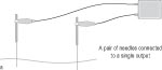
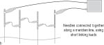
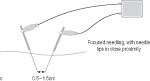
Brief LF EA (2 mA) may be more effective than brief moxibustion in prolonging latency of peripheral antidromic action potentials (APs), with EA mediated predominantly by substance P (SP), moxibustion by somatostatin (SS).359 Also, EA has been shown more effective than moxibustion in its inhibition of vibration-induced finger reflex.360 Apart from the obvious method of stimulating two separate AcPs with needles connected to an EA device, the circuit completed through the body (Fig. 6.2a), groups of needles can be connected, for instance along one or two meridians361 or in a localised area of pain,362 and stimulated from the same output (Fig. 6.2b). As an alternative method to treating two separate points, for strong local treatment two needles can be inserted close to and either side of an area of pain, so that although they enter the skin maybe 4 cm apart their tips are much closer together (0.5–1.5 cm) (Fig. 6.2c).363,364,365 Chronically implanted needlelike electrodes to stimulate otherwise inaccessible regions of the peripheral nervous system have been advocated in neurosurgery.366
| ||||||||||||||||
| To TENS or not to TENS? |
| |||||||||||||||
|
Clearly, in many respects EA and TENS are very similar in their neurophysiology, especially if used at the same locations. Some authorities believe they are virtually interchangeable when used at AcPs,367,368,369 although others emphasise correctly, if a little primly, that the term ‘electroacupuncture’ should not be used to describe treatments where needles do not puncture the skin.370 Much of the research on which TENS is based has been ‘poached’ from EA studies, especially the neurochemical ones. However, there are differences. For one thing, although surface stimulation can activate nerves even 4 cm beneath the skin,371 needle stimulation obviates problems of skin impedance (see SubCh. 5.1) so that much less current is needed to trigger action potentials (APs) in deeper afferent nerves than with surface electrodes (25 mA, with 1-millisecond pulse durations372). With the latter, at least with isolated pulses, there may be an initial transient current surge (due to the capacitative element of the impedance); this does not occur with needles.373 The deeper afferents contain a smaller proportion of thin unmyelinated fibres than superficial ones; such superficial C fibres may be activated by the higher currents needed with TENS.374 For these several reasons, EA may in some ways be less uncomfortable than TENS375 (skin/subcutaneous tissue PT tends to be lower than muscle PT, both being greater than fascial or periosteal PT376). However, in one case study comparing EA and TENS, although the pain relief obtained was similar, more urinary corticosteroids were found after EA, which possibly indicates that it was experienced as more stressful.377 (Patients may well be less apprehensive about non-invasive stimulation, even in China!378,379) The possibility that strong deep stimulation is endorphinergic and strong TENS less likely to be (see SubCh. 6.4, ‘gentle, shallow needling’) may also have some bearing on this. | ||||||||||||||||
| Studies where EA is more effective than TENS |
| |||||||||||||||
|
In a British study, local EA produced significant elevation of PT in response to cold immersion of the hand, whereas 100 Hz TENS did not. 8 Hz TENS also raised PT, but with considerable variation between individuals.380 In an important Japanese study of EA and TENS (both 100 Hz, 0.1 millisecond biphasic), only EA through needles insulated except at the tip significantly elevated PT in muscle and periosteum.381 Correspondingly, EA may reduce SEP amplitude more than TENS.382 Both EAA and TENS analgesia were significant in one study on low-intensity LF stimulation, but latency to EAA was shorter.383 Yoshiaki Omura reported that EA was more effective for wound repair than TENS, and also in reducing muscle spasticity when used at GB-21 (though TEAS was superior to MA).384 In rats with experimental diabetes, EA resulted in significantly lower plasma glucose, with attenuation of polyphagia, polydipsia and polyuria, raised PT and normalisation of motor nerve conduction after 4 weeks of treatment (20 minutes every 2–3 days, bilaterally at ST-36 and BL-23). In TENS-treated animals, plasma glucose levels reduced only slightly, motor nerve conduction normalised after 6 six weeks and PT decreased after induction of diabetes using streptozotocin i.p.385 Studies where EA and TENS are equally effective One major study by Han Jisheng’s Beijing group found that (1) both ST-36/SP-6 EA and TENS in rats produced analgesia of slow onset and offset at 2, 15 or 100 Hz (the time course being similar for both EA and TENS in each case), (2) systemic naloxone almost completely antagonised 2 Hz, partially antagonised 15 Hz and failed to affect 100 Hz analgesia resulting from both methods, and (3) tolerance to one form of stimulation at a given frequency reduced analgesia induced by the other form at the same frequency. The authors concluded that there is no significant difference between EA and TENS, in terms of either antinociception or the neural mechanisms involved.386,387,388 Richard Chapman also concluded that TENS and EA are pretty much equivalent.389,390 Low-intensity LF EA and TENS at ST-36 both resulted in analgesia and enhanced cellular immune function.391 In one Japanese study already mentioned, EA and TENS (both 100 Hz, 0.1 millisecond biphasic) significantly elevated PT in skin and fascia, while EA also (but not significantly) elevated muscle PT. The size, shape and material of the TENS electrodes (13 mm diameter rubber or metal cones, or soft rubber electrodes, 50 × 150 mm) did not greatly affect results.392 In further Japanese studies, both high-intensity TENS and EA (at 1 Hz, for 5 minutes) led to comparable increases in local skin blood flow, temperature and cold tolerance.393,394 One Chinese study found no significant difference between the use of EA or TEAS in the treatment of psychosis, although sleep improved more with the former. Deqi and PSM were reported by patients with both treatments.395 EA (5 Hz at BL-21, ST-36, Ren-12) and TENS (3 Hz) both significantly reduced gastric acid secretion in sham-fed humans, compared with MA or sham acupuncture.396 Meg Patterson found empirically that needles were unnecessary in her form of CES, and that pads were as effective.397 Studies where TENS is more effective than EA Han’s group, using a protocol of 2 hours of treatment every other day for 10 days, noted that tolerance to HF EA was greater than to TENS of the same parameters (100 Hz), especially by the fifth session (with reductions in both level and duration of EAA).398 Other Chinese researchers have found the analgesic effect of DD TENS greater than that of DD EA using similar frequencies.399 In one study on stressful stimulation, 20 minutes of noxious TENS (100 Hz, 0.1 ms, motor level, cathode to volar wrist, anode to upper arm400) resulted in localised reduction in experimental pain, but the same duration of noxious EA of ‘classical’ AcPs (88 Hz at maximum acceptable intensity, to LI-4 and points at anterior and posterior axial crease, radial and medial elbow crease401) did not produce any analgesia.402 Similarly, in a much quoted early study (small, and not placebo controlled), Andersson’s Lund group found that TENS increased dental PT more than EA despite almost identical subjective sensation and very similar onset and decline of effect.403,404 They considered that the larger electrode interface enabled higher currents to be used, resulting in recruitment of additional afferent fibres without excessive stimulation of nociceptive ones.405 One Japanese group found TENS more effective than EA for improving peripheral flap circulation and survival in the rat, possibly because stimulation was applied over a larger area in the former (the effects of intensity were unclear).406 | ||||||||||||||||
| Studies where MA is more effective than TENS |
| |||||||||||||||
|
In a study comparing MA, familiar to the subjects, with ALTENS (200 ms, gradually increased to elicit strong but non-painful muscle contractions at 15–40 mA), to which they were naive, dental PT increased significantly only with the former.407 MA at Du-26 in dogs was superior to TENS in increasing cardiovascular parameters under halothane anaesthesia.408 And in one small human study, temperature changes with HF TENS (albeit at 30–40 mA) were less than those found when testing MA.409 Studies where TENS is more effective than MA Both 2 Hz 40 mA and 100 Hz 20 mA TEAS at LI-4 increased the amplitude of the H-reflex evoked by soleus muscle stimulation in healthy volunteers, whereas MA at LI-4 for the same length of time (15 minutes) had no effect. Results suggest that TEAS enhances the excitability of the motoneuron pool in the spinal cord, but MA (at least, with the needle twisted initially to obtain deqi and then retained for 15 minutes) does not.410 | ||||||||||||||||
| Other comparisons |
| |||||||||||||||
|
EA (1–3 Hz, 0.4 ms, 10–12 mA at P-6 and (?) banmen (M-UE-13)) yielded a progressive, long-lasting and naloxone-sensitive decrease of the nociceptive blink reflex, whereas segmental TENS (80-100 Hz, 0.2 ms, 2-4 mA both at the wrist and at two points over the supraorbital nerve) had a more rapid effect that ceased immediately the painful stimulation was removed and was not affected by naloxone.411 Localised and general temperature changes induced by EA and TENS are not the same, although both may be sympathetically mediated (see SubCh. 6.3, ‘simultaneous sympathetic and parasympathetic activation’). In a study of low-intensity LF stimulation, naloxone partially antagonised EAA but not TENS analgesia, and whereas the former was almost completely blocked by deep (but not intradermal) injection of the local anaesthetic agent procaine, the latter was partially blocked by either deep or intradermal injection.412 Electrical stimulation of the median nerve produced a less detectable fMRI image from the contralateral sensorimotor cortex than when an acupuncture roller was used along the corresponding meridian (the image with rolling coincided with that obtained during hand and finger motor tasks, which was larger when these were more complex).413 In one TENS study, smaller (1.5 cm diameter) electrodes resulted in more local temperature increase/vasodilation than larger (4 cm) ones (electrode temperature did not rise); however, other parameters were covaried, such as frequency (3 Hz or 100 Hz) and intensity (1.5 or 3 times the sensory threshold of 5–9 mA), which makes interpretation difficult.414 Also, although electrode size, shape or material did not affect PT increases in the Japanese study mentioned above,415 or quality of sensation in one Italian report,416 others have suggested that effective stimulation may be experienced as unpleasant if electrodes are less than about 6 cm2 in area417 or 3 cm in diameter – at least using some waveforms.418 It certainly seems easier to obtain the electrical equivalent of deqi with such electrodes than with very small ones,419 presumably because more nerves can be activated (Ch. 4, ‘easier to activate more nerves’). With pads that are larger still, however, current density may be too low to activate deep muscle afferents.420 | ||||||||||||||||
| ||||||||||||||||
| ||||||||||||||||
| Other methods of stimulating acupoints and meridians |
| |||||||||||||||
|
Traditionally, moxibustion is a major part of acupuncture practice. There are many studies on its effects, for instance on rat TFL (comparing moxibustion at different AcPs and temperatures, needling with moxa, and different durations of heating426,427), on gastrointestinal electrical activity,428 on rat renal function and BP (using BL-15 and BL-27),429 on immune function and ageing (mouse thymus and pituitary activity,430 local immunocyte influx in rats and guinea pigs431 and spleen T-cell activity in mice432), on how different methods and dosages alter blood histamine levels433 or affect other neurotransmitters,434 on how it affects the EEG, enhancing alpha production,435 on intestinal activity436 and so on. The difference between heat applied through the needle (activating both muscular and cutaneous afferents) and radiant heat (activating predominantly cutaneous afferents) has also been explored.437 As an alternative to burning herbs, some ingenious electrical methods of heating have been developed. A method of ‘electrocautery’ of Du-26 using high temperatures (~80°C) for 10 minutes via a 3-millimetre probe significantly increased cardiac activity and BP in a number of studies on anaesthetised dogs under various circumstances,438,439,440,441,442 with more effect than EA, MA,443 TENS or finger pressure.444,445 Simple incandescent lamps have been used in many reports,446 or specifically infrared emitters.447 And many devices combine electrical heating with the use of herbs.448 One such even adds the benefit of an ‘electric wind’, claiming that a gentle low-temperature breeze for 10–20 minutes may strengthen wei qi (resistance to external disease), whereas a faster hotter breeze for 3–5 minutes (longer if directed at a more muscular area) may be used to treat febrile disease.449 The increase in PT that occurs with non-noxious heat (40°C) may be mediated by oxytocin (Ox) increases in CSF or plasma, or both.450 One study comparing moxibustion, incandescent heat and microwave concluded that there were no obvious differences between them (whether for xu or shi subjects), but that moxibustion instruments are more convenient to use than traditional methods.451 Another report suggested that combining EA with electrical warming of the needle might increase its therapeutic effect.452 Electrically heated needles have been used to stimulate immune function453 and in the local hyperthermia treatment of tumours (in mice, for instance454). Moist heat and shortwave diathermy (see Ch. 4, ‘27.12 MHz’) both relieved pain at trigger points (TrPs), with more effect from the latter at those that were only moderately sensitive initially.455 Not many studies have investigated the effects of cold (cryotherapy) at AcPs. Cooling the needle handle to induce small electric currents was suggested by a French doctor, Maurice Mussat, in the early 1970s.456 Recently, in an unpublished pilot study by British physiotherapist Lester Ward, a week of daily cold needling was found to reduce high levels of salivary cortisol possibly more effectively than using needles at room temperature.457 In a Chinese study, ‘freezing acupuncture’ was likewise found to increase T-lymphocyte counts significantly more than ordinary acupuncture in Kidney xu outpatients (most with chronic nephritis).458 Freezing acupuncture has been investigated for some respiratory conditions.459 Ice massage to LI-4 decreased acute dental pain significantly more than tactile massage (alone or accompanied by explicit positive suggestions) in a well-known study by Ronald Melzack and associates.460 Jeffrey Mannheimer and Gerald Lampe, in their classic tome on TENS,461 also refer to cooling AcPs. However, despite the existence of some Japanese equipment for cryotherapy of small regions, I know of only one study on a device for the relief of pain by cooling AcPs directly.462 In contrast, intermittent intense cold (with ice massage or a cooling spray) is a standard method for inactivating TrPs.463 There are some mentions of ‘microwave acupuncture’ (MWA) in the literature, although it is not always clear at first glance whether this is thermal or athermal in effect. MWA, for example, may (like EA) induce a temperature drop along a meridian from the treated point.464 Also, one comparative study found MWA analgesia to be greater than either MAA or that from HeNe laser acupuncture. 5HT was implicated in all three forms of analgesia.465 Millimetre waves (Extremely high frequency, EHF) This form of non-thermal stimulation was introduced in an earlier chapter (see Ch. 4, ‘Millimetre waves (extremely high frequency, EHF)’). One of its most frequent applications is in the treatment of peptic ulcer. For this condition it is used at AcPs (particularly ST-36466). A standard protocol would be 8–10 daily sessions, 20–25 minutes long, for example.467 Biopsies taken from 10 patients with duodenal ulcer showed an apparent acceleration of ulcer repair with EHF treatment.468 In rats (particularly those under stress), 60 GHz ST-36 EHF reduced gastric hypersecretion,469 and had both protective and remedial effects on experimental peptic ulcer (with 5- or 10-, but not 15-minute irradiation).470 In another human uncontrolled peptic ulcer study, EHF enhanced immune function,471 as it did in other conditions of compromised immunity when AcPs were irradiated.472,473 In one report,474 immune enhancement after five 20–30 minute treatments was dependent on the initial immune status of the subject, and could be reversed by overtreatment (> 15 sessions). EHF also resulted in a reversible increase of the sensitivity to certain antibiotics of some staphylococci at AcPs (although without any obvious direct effect on them).475 EHF at AcPs has been used in the treatment of experimental tumours in mice476 and rats.477 (Frequencies in the 35.9–42.3 GHz range, scanned at a 12.5 Hz rate, were more effective than in two higher ranges.) The effect of auricular EHF modulated at different frequencies on hypothalamic activity in humans has already been mentioned (see SubCh. 6.1, ‘local effect of EHF radiation on the brain’). Similar results for increased hypothalamic (and somatosensory cortex) activity in rats with SJ-20 55–65 GHz modulated at 26 Hz have also been reported,478 as well as effects (with different modulating frequencies) in rabbits.479 60 GHz ST-36 EHF may inhibit hypothalamo-pituitary neurosecretion and thyroid hyperfunction in rats, particularly those subject to stress.480 In humans, EHF (for instance, at GB-20, BL-10, SI-16 and SJ-13) is considered to reduce autonomic hyperactivity (whether sympathetic or parasympathetic).481 52–78 GHz EHF directed to a particular combination of body points (Ren-2, 6 and 18, Du-20, and bilateral BL-22, KI-3, P-6, LI-18) appeared to increase levels of plasma dehydroepiandrosterone (DHEA) in one American study using 3 minutes of daily irradiation in two 5-day courses followed by less frequent treatment.482 DHEA increases were more prominent if progesterone cream was also applied, and the Liss cranial stimulator (see above, ‘optimum parameters’) used too (!).483 In healthy volunteers, 52–78 GHz at Du-20 and HE-7, P-6, LI-4 and auricular shenmen (all on the left side) led to a general reduction in EEG power, particularly in the alpha (8–13 Hz) and beta1 (13–18 Hz) bands. Most decreases were greater on the left, particularly in the theta (4–8 Hz) band. Together, these effects are suggestive of greater relaxation. Changes were more marked in those unfamiliar with the treatment. However, control points were not used.484 In patients with chronic amputation stump pain, EHF to AcPs (classical or ashi points) reduced slow wave power in the EEG.485 In rabbits, low-intensity (10 mW/cm2) EHF at 55–75 GHz reduced hypothalamic EEG power in some narrow frequency bands, and enhanced it in others, with more effect at ear points than directly over the scalp. Exposure in both areas reduced power centred on 15.9 Hz, and increased that at 25.6 Hz.486 In studies on dental pain threshold in rabbits, equivalent results to those obtained with acupuncture were found with very-low-power EHF (1–20 mW); during this treatment breathing became more regular.487 Although LI-4 EHF (modulated at 1–10 Hz) was less effective than MA, it was more so than EA in this situation.488 Using skin potential (SP) measurement at AcPs (one on each of the Small Intestine, Kidney, Liver, Stomach and Spleen meridians on the left side) and non-AcPs, the return electrode being a needle inserted subdermally in the occipital region, EHF (70 GHz) and LF electrical stimulation (1 Hz, 50 µA) of the stomach meridian point (presumably ST-36) were found to have synergistic effects in dogs.489 EHF to the auricular ‘Heart’ point may reduce heart rate in rabbits.490 Athermal levels of EHF may have heating or cooling effects at AcPs outside the treated area.491 Subjective sensations of heat or cold, accompanied by corresponding changes in pulse rate, may occur too, as well as spreading sensations (PSM) of vibration or paraesthesia following a more local reaction, and even visual (colour) sensations.492,493,494 Intestinal peristalsis may also increase.495 Such ‘sensor reactions’ have been used to as a guide to optimal EHF wavelength,496,497 but may be less obvious in complicated cases498 and tend to become less pronounced with repeated treatment.499 The Omura ‘bi-digital O-ring test’ has also been used to select appropriate treatment parameters, which may well vary for the same patient.500 Locally, low-intensity (0.05 mW/cm2) 42.0 GHz EHF to P-8 leads to degranulation of dermal mast cells,501 as occurs too after EA (SubCh. 6.1, ‘degranulation of mast cells’). One authority on the use of EHF considers that reinforcement treatment should not be longer than 2–5 minutes, and reduction should be carried out for 15–25 minutes.502 In a very small pilot study, skin reflectivity to 61 GHz EHF at the AcPs P7, P-8 and P-9 was greater than that in neighbouring areas,503 and increased hydration at AcPs (in line with Hiroshi Motoyama’s findings (see SubCh. 5.2, ‘Motoyama Hiroshi’s acupuncture meridian measurements’) has been adduced as a reason why EHF may be better absorbed there than at non-AcPs.504 When the backs of rabbits (but not their eyes) were exposed to ordinary white light (but not yellow filtered light), continuous or intermittent (9 Hz), serum acetylcholinesterase significantly increased in the treated rabbits, but not in control animals.505 Blood glucose levels were affected quite differently by the continuous and intermittent light: they increased with the latter, but only well after irradiation.506 Intermittent light was also found to increase plasma dopamine.507 As with EHF, weak (athermal) infrared irradiation of the auricular ‘Heart’ AcP may alter both EEG and ECG characteristics.508 | ||||||||||||||||
| Low-intensity laser and polarised light |
| |||||||||||||||
|
Lasers and other forms of light with an energy density of around 40 J/cm2 or more will have a heating effect and may thus be comparable to moxibustion in some respects. Low-intensity laser (or light) therapy (LILT) is quite different, as discussed above in general terms (see Ch. 4, ‘Low-intensity lasers and polarised light’). LILT in the form of ‘laser acupuncture’ (LA) has in particular been promulgated as a pain-free and non-traumatic alternative to needles509 or even TENS.510 Invasive LA is also used, the light being conducted via an optic fibre threaded through a hollow needle, so combining the benefits of LILT and acupuncture (or even EA511). In terms of the very different mechanisms involved, the term ‘laser acupuncture’ should perhaps be restricted to the invasive form,512 but here ‘LA’ will be used to cover LILT as applied cutaneously to AcPs (if needles are used, this is stated explicitly). There are, as yet, few studies of the effects of LA within the brain, but in one pilot study diode LA (880 nm, 1 J/cm2) at Du-14 and TrPs in the trapezius muscle was found to alter regional cerebral blood flow significantly in various parts of the brain associated with nociception (the thalamus, caudate nucleus and prefrontal cortex).513 The effects of LILT on pain are still controversial (see Ch. 4, ‘More controversial than the use of LILT’). In particular, the opioid effects of LA are still not agreed. For instance, blood levels of βEP and LE may be normalised using HeNe LA.514 In keeping with this result, i.v. naloxone was found to reverse PT increases in rabbits in one GaAs ST-36 LA study. Here AcP irradiation had very similar effects to deep fibular nerve irradiation, suggesting, in contradiction to Thomas Lundeberg,515 that LILT may indeed owe its effects to nerve stimulation.516 However, in rats the increase in PT obtained with 4 Hz HeNe LA at Du-1 was not reversed by a low dose of naloxone (2 mg/kg) given beforehand. In this study, PT changes were assessed by TFL and hot plate (HP) methods, and it also transpired that a high dose of naloxone (20 mg/kg) did actually reverse the LA effect for HP but not for TFL.517 Naloxone was also unable to block TFL increases in a further study, using 60 Hz pulsed LA.518 However, tail flick is a local spinal reflex (unlike HP), not centrally mediated, so really this is hardly surprising. A RCT of auricular HeNe LA in healthy subjects demonstrated a significant increase in PT (to painful electrical stimulation of the ipsilateral wrist) in healthy subjects, with no such change in the sham treatment group,519 despite a low-dosage protocol that was subsequently criticised by David Baxter (as was use of PT as the sole outcome measure).520 Baxter also expressed concern about the dosage used in a SEP study of HeNe LA (0.7 mW or 1.26 J at unilateral LI-4 and P-6 for half an hour) in which effects faded after an hour,521 suggesting that results might be due to expectation and habituation rather than LA per se. However, the author of this last study cites another Chinese study in which SEP effects were similar with both LA and ‘needle acupuncture’ (presumably MA).522 In one Chinese study, though, the effect of ST-36 HeNe LA on pain tolerance (rather than PT) was found to be a useful pretest for the epidural dosage required for gastrectomy when the epidural was used in combination with AA.523 Some reports give more clearcut results on LA and pain. In 10 out of 14 horses, infrared LA at nine AcPs (300 mW, 904 nm, 360 Hz, for 2 minutes per point) significantly alleviated signs of chronic back pain and greatly improved performance (with continuing benefits 1 year post treatment).524 In rats with experimental hindpaw arthritis, HeNe BL-60 LA (3 mW, 10 minutes daily for 5 days) significantly reduced ankle swelling and pain scores more than sham or no treatment (but without any effect on PT except immediately after treatment).525 Also, LA significantly increased PT in rabbits, dogs, pigs, sheep and goats, the effect being potentiated by administering a combination of L-tryptophan, D-phenylalanine, D-leucine, acetylcholine and insulin (!).526 This is in line with findings that 5HT, OPs, ACh and other neurotransmitters may mediate LILT (see Ch. 4, ‘LILT may significantly alter brain levels’), and the effect of hypoglycaemia on EAA (see below, ‘hypoglycaemia may potentiate ALS’). LA may have cardiovascular effects. Thus HeNe laser irradiation of P-6 in cats (using 2.7 J/point) has been shown to reduce HR, with other effects on the ECG as well.527 HeNe LA has similar effects in cows, together with a reduction in haemoglobin levels (the same changes, and some additional ones, were observed if the uterine cervix was irradiated, rather than AcPs528). However, in rats HeNe LA (or associated stress?!) may increase HR (and physical activity in general, with decreased AChE particularly in young rats).529 LA also influences ventricular function in humans (3–5 mW HeNe at P-6).530 However, although P-3 LA speeded recovery in rabbits with acute myocardial ischaemia (AMI), it did not reduce myocardial injury if used after severing afferent nerves from that point, which indicates the involvement of the afferent pathway in LA.531 LILT is well known for its effects on tissue repair. HeNe LA, in combination with the bioflavonoid quercetin, has been shown to reduce postoperative inflammation of injured nerve.532 HeNe LA also improved microcirculation and enhanced muscle regeneration in rats with experimental myopathy due to denervation,533 particularly when combined with physical loading.534 Myopathy due to forced inactivation (including an increase of slow muscle fibres) was inhibited by LA.535 As with EA, VIP and CGRP have been invoked to explain the tissue repair effects of LA.536 LA may have particularly useful effects on the immune system. It appears, for example, to enhance some cell-mediated and humoral immune characteristics in rats537,538 and mice, as well as non-specific antiviral resistance (to acute experimental influenza) in the latter.539,540 Bilateral 2 mW HeNe ST-36 laser irradiation (10 minutes daily, 14 treatments in all) may beneficially affect a number of immune parameters.541 However, the effect of using ‘photobustion’, an incandescent light flashing at 0.5 Hz, to heat points to 40–43°C daily for 10 minutes, was not significantly different. Direct irradiation of the thymus area in rats exposed for the first time may induce a stress response. Rather as with EA (see SubCh. 6.4, ‘greater increases in serum cortisol’ and ‘relaxation only in subsequent sessions’), repeated (or longer) treatment may reverse this, but with little effect on immune parameters.542 However, some effects of LA such as those on chronic inflammation may well be cumulative.543 Laser cautery, like the use of strongly heated needles, may elevate leukocyte counts and serum immunoglobulins.544 Invasive LA has been used to irradiate the blood directly as a means to enhancing immune function.545 LI-20 HeNe LA (2.5 mW) has been used in the treatment of experimental allergic rhinitis, resulting in temperature increases of up to 5°C at the contralateral LI-20 point.546 Rather as with EHF (see above, ‘Such ‘sensor reactions’’), sensor reactions of warmth, tingling or sharp pricking may indicate that treatment has had an effect and need not be continued.547 Such deqi, if one may call it that, is perhaps similar to that obtained with MA, but usually much less marked.548 However, in one informal report of 450 treatments using the LASERneedle system (see Ch. 11), deqi was obtained in over 90% of cases and was found to be greater than with MA, particularly in acute cases.549 In one rabbit study, however, ST-36 HeNe LA did not inhibit anaphylactic reactions, although HeNe LA did appear to have some regulative effect on blood histamine levels.550 LA affects gastroelectric activity in rabbits551. Also, Du-1 LA has been used for dysentery in young lambs, with rapid reduction of hyperperistalsis and borborygmus.552 Polarised light (together with MA), which is associated with partial recovery of sensation, has been used on scalp and back acupoints in a dog with loss of sensation following a road traffic accident.553 Comparative studies of ‘laser acupuncture’ Some careful studies have been carried out comparing laser and acupuncture for pain. In one on rats by Thomas Lundeberg’s group,554 using HeNe and GaAs lasers at the base of the tail, both power (1.56 and 0.07 mW) and energy (~1.4 J and 0.06 J per point over 15 minutes) were probably too low to be effective. So although 80 Hz local EA did prolong TFL, the conclusion that LA analgesia might be due to placebo is unwarranted. In a German study, single-blind MA (bilaterally at LI-4) increased PT to painful heat stimuli more than MA at a control point, and also more than double-blind HeNe laser irradiation for 1 minute at each point (bilaterally at LI-4 and jianqian, M-UE-48).555 However, this study has been criticised by David Baxter as depending solely on PT as the outcome measurement, and (like studies by Lundeberg’s group) because the laser dosage used was very low.556 Ukrainian researchers (using non-specified LILT parameters) found no analgesic effect with LA.557 Yet Chinese researchers found that 30 minutes of either EA or LA increased amplitude of the ‘N80’ component of SEP in response to median nerve stimulation, although it look longer for the N80 to return to pretreatment level with EA (20 minutes) than with LA (10 minutes). Importantly, they found that there was no effect on N80 amplitude in normal subjects558 – something that might help to explain the negative results of some LILT nerve conduction studies (see Ch. 4, ‘enhanced slow components’). In a Hong Kong study, both LF EA (20 minutes, intermittent) and LA (0.6 J/point, 120 seconds per point, ‘closed contact’ method) at LI-4 and ST-36 decreased late component (cognitive) SEP amplitude in response to painful dental stimulation. Results using EA were marginally superior to those using LA.559 In a study of chronic back pain in horses, LA (904 nm, 300 mW, 360 Hz, for 2 minutes/point), MA (even needling) and ‘injection acupuncture’ (with isotonic saline) all appeared to be equally useful.560 Similarly, TENS, EA and LA all gave comparable results in one human case study.561 HeNe laser at ST-36 resulted in similar levels of analgesia and enhanced cellular immune function to those obtained with TENS or EA (low-intensity LF) at the same AcP.562 A further comparison, between HeNe ST-36 LA and ‘photobustion’, was mentioned above.563 When compared with MA, HeNe LA was found to elevate PT significantly more than MA in rabbits, with (infrared) CO2 being more effective than HeNe LA in dogs.564 However, in rats, lower power HeNe LA resulted in greater analgesia in one study (6 mW as opposed to 15 mW), LA at both powers being less effective than MA and MWA.565 Yoshiaki Omura reports that 15 minutes of 10 mW GB-21 LA was less effective in reducing muscle spasm (cramp) than EA, TENS or even MA.566 Litscher and Wang, in a small pilot study, found that the effects of LA (785 nm, 19.6 mW) on cerebral oxygenation were less than those of manual needling for the same length of time (20 seconds). With both modalities, effects were greater at the acupoints used than at a sham point.567 LA and VLF low-intensity EA at P-6 have been compared for their effects on cardiac function. LA appeared to have greater effects in this study.568 In a single case report in which various stimulation modalities were used, urinary glucocorticoids increased more when LA was pulsed at 2 Hz than at 64 Hz.569 In a more detailed study, 1 mW HeNe laser at low SR tail points (~Du-1) in rats for 15 seconds resulted in rapid onset short duration (24-hour!) hypoalgesia when pulsed at 4 Hz, delayed and longer lasting (48-hour) effects on TFL at 60 Hz, and no effect at 200 Hz.570 (Compare the Ulster study on 16 Hz versus 70 Hz mentioned above, see Ch. 4, ‘slower in onset yet longer lasting’.) As David Baxter comments, this appears to be a genuine frequency effect (as power differences at the different frequencies were taken into account). Less clear is a result reported by Jean-Claude Darras: 28 mW HeNe LA tended to affect the rate of flow of a radioactive tracer through the connective tissue more when pulsed at 24 Hz than 48 Hz (but the method of pulsing was undefined).571 LILT was found to be less effective than both 3 Hz EA and 5 Hz TENS in reducing sham-feeding-induced gastric acidity in humans.572 Preliminary data from an Australian group indicate that near-IR laser (780 nm, 3 mw) is preferentially absorbed when pulsed at LF (50/50 duty cycle) by the AcP LI-4 compared with a non-AcP (both located by measuring SR). Above 10 Hz, no difference was found.573 Although this study is important in that it draws attention to a possible link between low SR (thin corneal layer? greater hydration?) and LILT absorption, and may shed some light on variation in response using different pulse frequencies, it is not entirely clear whether the result is in fact due to the different frequencies used or to cumulative effects with longer pulse durations. | ||||||||||||||||
| Low-intensity pulsed electromagnetic fields |
| |||||||||||||||
|
Very low (pTesla) PEMF applied to the head may increase release of 5HT or enhance 5HTergic transmission.574 Stronger fields (on their own, or in combination with CES) may induce drowsiness.575 In rats with depressed humoral and cellular immune function (following lesions to the anterior hypothalamus or caudate nucleus), magnetic stimulation may to some extent restore immune function.576 In a small pilot study, Saul Liss noted trends to increased blood levels of 5HT, βEP and ACTH, with decreased cortisol, after treatment with the Medicur device (see Ch. 11).577 While PEMF has been applied to TrPs in clinical practice,578 there appear to be few experimental studies on PEMF within the context of acupuncture. Small magnets (750 or 1500 G) applied to AcPs may enhance leukocyte activity, phagocytosis and a non-specific immune response.579 Magnets applied to the skin, or even light finger pressure in the presence of a small (2 G) magnetic field, may reverse, deviate or weaken PSM induced by LILT.580,581 In rabbits, a 1300 G magnet (‘North pole’) applied over muscle may restore muscle twitch depressed by long-term tetanic stimulation (10 Hz, for 1 hour), possibly by re-establishing local microcirculation via cholinergic mechanisms (i.e. inhibition of AChE). This might well have an effect on local muscle pain as well.582 The use of magnets in conjunction with Yoshiaki Omura’s bidigital O-ring test is mentioned below (see below, ‘magnets are positioned’). Jacques and Andrée Pontigny have suggested that magnets may be beneficial only if the subject is moving relative to the geomagnetic field; wearing them at night may thus not be useful.583 Pain relief with 100 Hz vibration is not affected by (low-dose) i.v. naloxone.584 However, non-noxious 100 Hz vibration may be mediated (at least in rats) partly through activation of oxytocinergic mechanisms.585 100 Hz vibration may also induce vagally mediated increases in plasma gastrin and cholecystokinin (CCK) levels, as may 40°C warming or 2 Hz EA, with SS changing only in response to the latter, that is intramuscular stimulation.586 Furthermore, adenosine appears to mediate HF (100 Hz) vibration-induced analgesia,587 as well as low-intensity HF TENS analgesia or EAA (see SubCh. 6.2, ‘two or three cups of coffee’, and SubCh. 6.4, ‘mediated by adenosine’). 100 Hz vibration and TENS, as well as 2 Hz EA, have all been found superior to placebo in the treatment of myalgic pain.588 Like TENS and EA, 100 Hz vibration does not affect thermal sensitivity to non-noxious heat, even if relieving clinical pain in the same subjects.589 When used intrasegmentally at or proximal to a site of experimentally induced pruritus, vibration at 10 Hz, 100 Hz or 200 Hz (and 2 Hz or 100 Hz TENS) reduced subjective itch intensity, with 100 Hz being the most effective especially locally. Itch duration also decreased most after prior 100 Hz vibration, which had the most rapid effect as well.590 However, 100 Hz vibration and TENS had no effect on experimentally induced itch when applied extrasegmentally, although 2 Hz TENS did.591 In contrast, some researchers have noted that vibration, at 60 Hz for instance, does have contralateral effects on itch592 or, at 20 Hz, 50 Hz, 100 Hz or 200 Hz, on pain.593,594,595 Clearly, 100 Hz vibration stimulating large-diameter cutaneous afferents shares quite a number of characteristics with HF TENS or (gentle) EA, even though these may activate Aβ or even Aδ. Generally, vibration at > 50 Hz takes effect rapidly and that at < 10 Hz more slowly.596,597 The effects of 20 Hz vibration, however, are mixed: as with LF EA, the increase in PT is slower in onset than with HF stimulation but, as with HF EA, more rapid in returning to pretreatment level. Importantly, with 20 Hz or HF vibration the duration of effect on clinical pain may be much greater than that on experimental PT. Increases in PT occur in phase with enhanced local (and possibly diminished peripheral) blood flow.598 LF vibration of the acupuncture needle is sometimes used in MA to good analgesic effect.599 In rabbits, twitch response to stimulation at TPs was greater with vibration (tapping) than simple ‘dry needling’ (though less than with the mechanical equivalent of snapping palpation).600 Although sound has been applied to AcPs in clinical practice,601,602,603 there are few experimental studies in this area. Some research has been carried out on the ‘Qi Gong machine’ (see Ch. 10). In a more traditional vein, Susan Tomkins, an acupuncturist trained in the Five Element approach, has looked at the effects of 329.6 Hz applied to Earth points on the different meridians.604 Despite the paucity of research, some devices for audiofrequency electrical stimulation have been utilised.605,606 Clinically, the combination of sonic with electrical stimulation may markedly affect body temperature.607 As already mentioned (see SubCh. 5.2, ‘may also have bioluminescent’), meridians may have their own acoustic characteristics. Very-low-intensity ultrasound (0.05–0.2 W/cm2) has been used to treat auricular AcPs (even at 0.4 W/cm2, repeated ultrasound stimulation could lead to electrical ‘hyperactivity’ at the points).608 In one controlled study, 20 minutes of ultrasound applied to ST-36 was found to increase somatic and gastralgia PT in rabbits, the effect peaking 25–30 minutes post treatment.609 Use of a special applicator to ‘focus’ ultrasound deep within the tissues at ST-36 and ST-41 also improved microcirculation in the human leg.610 Ultrasound has been used too to detect TrPs611 and to investigate differences in sensation at body and auricular AcPs.612 In one fascinating study, Joie Jones (a colleague of Zang-Hee Cho at UC Irvine) found that athermal 50–80 W/cm2 ultrasound, at various frequencies (centred on 5 MHz) and applied through a 2 cm diameter head mounted in a water-filled flexible chamber over BL-67, resulted in fMRI records of occipital lobe activity indistinguishable from those obtained with BL67 MA (see too SubCh. 6.1, ‘activation of the visual cortex’).613 Optimal results were obtained when the ultrasonic pulse was focused at the same depth as the needle tip had been; if focused more deeply or on the skin surface, no cortical activity was observed. | ||||||||||||||||
Possible adverse effects of stimulation |
| |||||||||||||||
EA has endorphinergic effects, with the implication that awareness of painful stimuli may be dulled, possibly resulting in injury. However, as Anders Ekblom and Per Hansson found, while EA may reduce clinical pain intensity, thermal sensitivity (for instance) is not affected.614 This has also been reported for SPES615 and some other forms of electrostimulation.616 However, during TENS treatment, and for a short while after, sensitivity to electrodiagnostic testing (the rheobase, see Ch. 4) may be elevated.617 With EA, if a nerve is slightly injured by an acupuncture needle the threshold to subsequent stimulation may be reduced, so that EA may be experienced as more painful than usual until the nerve has healed.618 In general though, even with repeated treatments, there do not appear to be any long-term adverse effects on nerve conduction characteristics with peripheral electrical stimulation.619 If chronic pain is the result of severely impaired descending inhibitory pathways, then EA or TENS may exacerbate rather than relieve it.620 This aggravation might provide a clue that such impairment is present, and could well be less likely to occur when using methods such as CES that do not directly affect primary afferent synapses in the dorsal horn. Remembering the Arndt–Schulz law, excessive levels of stimulation (whether in terms of amplitude, or pulse or treatment duration) may be counterproductive621 – not least because they may trigger a stress response (see SubCh. 6.4, ‘stress mechanisms became involved’). Stressful stimulation may adversely increase plasma/blood viscosity and erythrocyte aggregation rates, for instance.622 Strong (or prolonged) stimulation should be avoided at particular AcPs such as Du-3.623 Somebody who is already stressed may experience even low-level stimulation as stressful, with possible adverse responses.624 Strong electrical muscle stimulation (as opposed to comparable voluntary contractions) may temporarily damage muscle fibre membrane.625 As with any treatment that leads to muscle movement, motor level EA or TENS may affect tissue uptake or distribution of locally or systemically administered substances. While this effect may be usefully harnessed, it is not always desirable. Spinal cord stimulation may reduce heart rate via sympathetic inhibition.626 This should be remembered when using dumai (Governor Vessel) or huatuojiaji points in yang xu patients. HR may also be affected by DC stimulation of HE-9 with Ren-17 (see SubCh. 6.4, ‘adverse, if temporary, cardiac effects’). Vagal stimulation in rats may increase the proportion of degranulating mast cells in the trachea and bronchi (more so on the stimulated side), mediated possibly by polymodal neurons of the jugular ganglion.627 If a similar pathway exists in humans, auricular stimulation (especially bilateral) might be expected to cause bronchoconstriction. Caution is generally advised in stimulating points on the throat, in part because of possible baroreceptor effects via the carotid sinus, leading to HR and BP reduction, and in part because such treatment might lead to airway restriction. However, using TENS (1 ms, 2.5–4 mA) both unilaterally and bilaterally over the tracheoesophageal groove led to increasing glottic aperture at lower frequencies (maximal at 30 Hz) and closure only at higher frequencies (total closure at 100 Hz), with no effects on respiration or heart function.628 Certain stimulation frequencies have been considered to be in some way undesirable. For example, if HF EA or TENS releases spinal cord dynorphins, and these inhibit vasopressin and LHRH (SubCh. 6.3, ‘Gonadotropin-releasing hormone (LHRH or GnRH)’), it is possible (if unlikely) that such stimulation might inhibit or delay ovulation (see SubCh. 5.1, ‘dynorphin may inhibit release’). LF stimulation that leads to 5HT-mediated peripheral vasodilation can also sometimes result in headache.629 | ||||||||||||||||
As a generalisation, symmetrical biphasic waveforms produce more effective stimulation and fewer side-effects than others.632 Although DC EA633 and EA hyperthermia have both been used in the treatment of tumours (see too Ch. 4, ‘Rudolf Arndt, a psychiatrist’), they are very much experimental procedures, not a part of normal clinical practice. If poorly understood, it is very possible that they might enhance tumour growth or metastasis. However, there is no indication that very-low-intensity stimulation (such as SPES) can adversely affect cancer,634 and one report on the use of EAA (MAA?) with epidural anaesthesia in a small number of patients (14) found no evidence of any lymphocyte mutagenic or carcinogenic effects.635 Nevertheless, metastatic activity was dramatically increased in one study of rats receiving 100 Hz PAG stimulation analgesia after injection with carcinosarcoma (ascites) cells.636 In view of some similarities between this form of analgesia and EAA, it would seem sensible to err on the side of caution when treating patients known to have cancer. Of course, in general clinical practice needles should not be used with DC stimulation because of the effects of electrolysis637 (see Ch. 3, ‘thoroughly explored’). Even without overt electrolysis, needles may undergo chemical reactions at the metal surface, with the possible adsorption of residual byproducts.638 Thus they should not be reused, certainly without proper cleansing and preferably not at all.
An in vitro study on the hippocampus from young rats (< 2 weeks) shows LF stimulation may have double the effect found in that from older ones (> 5 weeks).643 As the effects being studied (reversible long-term depression and potentiation of neuronal activity) are very probably also involved in the dorsal horn synaptic mechanism of EA and TENS, this is a pointer to caution in the treatment of children. Furthermore, if there is any substance to some of the theories of EEG resonance described in the next chapter, the effects of LF repetitive stimulation on children whose brains do not yet show adult patterns of activity may be quite different from those found in adults. For instance, in children there is a more ‘introverted’ dominance of theta rather than of alpha, as in adults.644 Until such differences are clarified, caution is again indicated. | ||||||||||||||||
LILT | | |||||||||||||||
Although dosage, at least for tissue repair, should be ~4 J/cm2 (Ch. 4, ‘~4 J/cm2’), HeNe even at 0.3–0.5 J/cm2 was observed to cause considerable damage to the mitochondria of fruitfly (Drosophila auraria) larvae, but not at < 0.3 J/cm2.645 Whether this is significant in terms of human mitochondrial damage is another question. Caution should be observed when using LILT on a patient who is taking St John’s Wort (SubCh. 6.2, ‘cautiously in conjunction with LILT’). | ||||||||||||||||
|
| ||||||||||||||||
Strong visual stimulation may induce seizures at certain low frequencies (12–18 Hz),646 though not at 4 Hz for instance,647 unless long pulse durations are used.648 This effect is probably also wavelength dependent,649 white or blue light being safer than green or red, for instance.650,651 However, the risk of triggering epilepsy is minimal with LILT, for which in any case safeguards should be in place to prevent direct exposure of the eyes during clinical use. In patients with physically obvious pre-existing exaggerated responses (as in myoclonus) or abnormal EEG patterns, prolonged nerve stimulation at particular frequencies (in one case at 10–14 Hz, for example) may lead to generalised convulsions. Fortunately such patients are not commonly encountered, and are able to recognise something different and uncomfortable in the overall subjective sensation elicited by stimulation before convulsions occur.652 Early reports of this epileptogenic effect of LF stimulation were used by Meg Patterson to support her use of HF CES,653 but since then it has been found quite unlikely that even direct visual stimulation at < 15 Hz654 or magnetic stimulation at < 17 Hz655 will cause problems, while the extrapolation to somatosensory stimulation may be quite unjustified for the vast majority of patients. For one thing, epileptogenic ‘kindling’ (see Ch. 7) is more likely to occur at higher frequencies, even when the brain is directly stimulated.656,657 Although LF (3 Hz) amygdala stimulation has been used to induce kindling in rats,658,659 for example, elsewhere within the brain LF (1 Hz DC or 4 Hz biphasic) may ‘quench’ such amygdala-kindled seizures.660,661,662 Secondly, generalised seizures tend to occur only in patients with a personal or family history of epilepsy,663 and kindling is unlikely to occur in those without such a history.664 However, any sign of rhythmic myoclonic jerks occurring in time with the applied stimulation (or at a harmonic of that frequency), particularly in such a patient, should be viewed as a contraindication to continued treatment (mild local muscle twitching in response to EA or TENS occurs commonly, and should not be confused with myoclonus). Thirdly, EA has been found to reduce experimental epilepsy and convulsions, and their effects in the brain, in a number of animal studies,665,666,667,668,669,670,671,672,673,674,675,676,677,678,679,680 although in one detailed report on work with cats and mice the results were not so clearcut.681 MA or EA (2 Hz trains of 50–70 ms, internal frequency 950 Hz, 0.2 ms, intensity not indicated) was applied to 24 acupoints used clinically for epilepsy, in various combinations. In another study using MA at the same points (some distally on the limbs, some on or near the head, such as GB-20, Du-14 and Du-26), acupuncture clearly increased EEG spike activity in most animals, particularly if used during seizure discharges.682 Prior MA has also been found to reduce induced epileptiform EEG in rats.683 Rats prone to seizures (with a functional defect in central CCK mechanisms) may in fact respond better to EA than other rat types in some respects.684,685 EA at ST-36 has been used to inhibit cAMP-induced epileptiform waves (in amplitude, frequency and duration),686 and at Du-26 and Du-24 to prevent hyperbaric oxygen-induced convulsions.687 Lumbar points on the dumai (Governor Vessel) seem to be particularly effective, tending to inhibit both amplitude and frequency of epileptiform hippocampal activity, for instance, especially at higher intensities, with frequency (1 Hz or 5 Hz) seemingly a less important factor.688,689 However, MA at non-acupoints that do not effectively inhibit epileptiform activity may in fact potentiate it (see SubCh. 6.1, ‘potentiated epileptiform activity’), and repeated treatment may reduce antiepileptic effectiveness (possibly through enkephalin tolerance).690 Furthermore, strong EA applied to head points may occasionally exacerbate penicillin-induced epileptiform bursts691 and MA, as mentioned, may also increase penicillin-induced seizure activity.692 The antiepileptic effects of EA may be mediated by 5HT.693,694 GABA has also been implicated in Du-24/26 attenuation of hyperbaric convulsions695 and, with endorphins and CCK, in penicillin-induced epileptiform discharges.696,697,698,699 Hippocampal βEP and LE elevated by ECT or penicillin microinjection are reduced by protective EA (134 Hz, 7–10 mA) at Du-8 and Du-16,700,701 probably as a result of it down-regulating their synthesis (in this study EA at the same points was at 100 Hz, 6 mA, so still an intense stimulation).702 However, Enk may have both pro- and antiepileptic activity.703 Dynorphin levels, in contrast, which are reduced after penicillin-induced seizure, may be enhanced by EA.704 The anticonvulsant effect of EA may also be mediated by decreases in some NO synthases.705 Clinically, auricular acupuncture (with press needles) has been used for canine epilepsy,706 and EA may have useful antiepileptic effects in humans707 (see SubCh. 9.1). SPES too may have some antiepileptic effects.708 Nevertheless, as epilepsy may involve repetitive discharge with reduced variability in some parts of the brain that are also involved in acupuncture pathways (see Ch. 4), the use of regularly repetitive strong stimulation should be avoided particularly at points near or on the head. As has been shown repeatedly, high-intensity ‘electronarcosis’ like ECT may well induce convulsions.709 HF EA at 7–10 mA has been used to attenuate such convulsions in animals710 (decreasing β and increasing δ band EEG activity711), but also, at even higher intensity, to induce convulsion as a less damaging alternative to ECT.712 Strong EA used for prolonged periods in surgery has likewise been found to trigger seizures.713
A number of drugs may induce epileptiform activity in the brain. These include chlorpromazine and lithium (markedly), thioxanthenes, butyrophenones and amitriptyline (moderately), and imipramine, monoamine oxidase inhibitors (MAOI)s, thioridazine, meprobamate, chlordiazepoxide and methylphenidate (slightly).717 This is more likely to occur during the first weeks of treatment or during withdrawal (as may occur too with drugs such as haloperidol that lessen seizure frequency if taken for long periods718). EA should be used with particular caution during these periods. Even during alcohol withdrawal, although spontaneous epileptiform activity is rare, a ‘photomyogenic’ response may be triggered by photic stimulation. Bearing in mind the similarities between photic and somatosensory stimulation (Ch. 7, ‘cooperativity between neurons’), use of EA at this time should be undertaken with caution. Cocaine use can lead to seizures,719 and amphetamine may sensitise to subsequent kindling,720 so the possibility of adverse effects from EA in past users should not be overlooked. Prior exposure to some pesticides may also predispose to seizures.721,722,723 However, regular exercise may inhibit the development of kindling.724 | ||||||||||||||||
| ||||||||||||||||
Because acupuncture can sometimes lower temperature,725 it has been suggested that care be exercised when using strong needle manipulation in yang xu patients.726 However, with comfortable (non-stressful) EA, which in general is warming (SubCh. 6.3, ‘generalised temperature increase’), this caution may not apply. Chronic or acute stress may interfere with 5HT metabolism, and so with many forms of stimulation.727 Anxious patients may be less able to tolerate electrical stimulation,728 and anxiety in general is found to reduce EA effectiveness.729,730,731 Paradoxically though, it may sometimes result in greater PT elevation.732,733,734 Patients with low initial PT may respond better to EA than those whose PT is already high.735 However, in those with neurogenic pain (e.g. radiculopathy), LF electrical stimulation of the involved nerve can sometimes cause the pathological pain to be reproduced, if temporarily.736 As mentioned above, exacerbation of chronic pain may occur if descending inhibitory pathways are impaired in some way. In diabetic rats, the analgesic potency of morphine is reduced,737 probably due to changes in morphine’s kinetics rather than opiate receptor characteristics,738 although direct PAG stimulation does appear to be mediated by different opioid receptor interactions than in normal rats.739 Thus ALS, like morphine,740 may be less effective at ‘low doses’ when it comes to clinical practice. However, diabetes does not appear to alter the potency of analgesia produced by delta- and kappa-opioid agonists,741 so that HF electrostimulation is less likely to be affected. Interestingly, it is possible that hypoglycaemia may potentiate ALS, as it does the antinociceptive effects of aspirin and morphine (at least, when insulin-induced).742 A study was also mentioned above in which insulin potentiated LA (above, ‘acetylcholine and insulin’). One implication of these results is that EAA and also LA effects may be reduced in diabetic human patients. Parkinson’s disease, which decreases cerebral 5HT, might reduce the effectiveness of EA and related methods.743 Patients with recent cerebral haemorrhage should be treated cautiously, owing to the vasodilatory effect of acupuncture.744 In rats, opioid receptors may decline during ageing, which may also increase hormonal responses to stress.745 It may be necessary to take both these factors into consideration when treating elderly patients with EA. Decline in pineal function with ageing may also reduce opioid-mediated analgesia.746 However, ST-36 EA (2 Hz, 1.5 V) was found to facilitate sensory transmission to S1 (in rats where it had been strongly inhibited by β-amyloid topically applied to S1 as a model for the neuronal alterations in early Alzheimer’s disease), although surprisingly it did not modulate this transmission in normal rats.747 Adequate nutrition is a prerequisite for SPES748 and presumably to some extent for EA too: without sufficient precursor intake the neurotransmitter mechanisms involved may not be able to function efficiently, as very readably described in the book Natural Highs by Patrick Holford and Hyla Cass.749 Vitamin B6, for instance, may facilitate some GABA- or 5HT- mediated EA effects.750,751 In some circumstances, as mentioned (above, ‘hypoglycaemia may potentiate ALS’), hypoglycaemia may potentiate the effects of ALS. Furthermore, hypoglycaemia (e.g. due to fasting) may exaggerate EEG anomalies and so potentially also anomalous responses to EA (particularly in those with a personal or family history of epilepsy).752 However, food intake shortly before acupuncture treatment may affect both breathing rhythm and peripheral blood flow,753 which may be misleading, for instance, to a TCM practitioner. (This section can usefully be read in conjunction with Table A3.1 in Appendix 3) Any strong stimulation to the head in a patient on anticoagulant drugs should be undertaken only with great caution, because of its potentially vasodilatory effect (this is an extrapolation from findings with ECT, electroconvulsive therapy).754 This may even apply to those taking such seemingly innocuous drugs as aspirin, or alternatives such as gingko biloba or garlic. Caffeine (and theophylline), which is found in coffee, tea and cola drinks, may reduce the effectiveness of HF stimulation, whether EA, TENS or vibration, through antagonistic effects on adenosine755 (SubCh. 6.2, ‘two or three cups of coffee’). Caffeine may also affect peripheral blood flow and breathing rhythm,756 as well as increase circulating βEP.757 Foods or beverages containing these stimulants should therefore be avoided for some hours prior to HF (and probably LF) EA treatments.758 However, caffeine does not adversely affect SPES.759 Any drug that enhances spontaneous EEG rhythms within a particular frequency range may potentiate the effect of strong EA or TENS at similar frequencies.760 Monosodium glutamate (MSG) may adversely affect SPES if the blood/brain barrier is leaky,761 and also EA762,763 (presumably in the same circumstances). Because it selectively affects the hypothalamic NArc (see SubCh. 5.1), MSG is more likely to reduce the effectiveness of CEA than TLEA.764 Whereas chronic L-DOPA (for Parkinson’s disease) may deplete 5HT (and so interfere with EA), the antidepressant sertraline, which inhibits 5HT reuptake and is often given for Parkinson’s, may improve response to EA and allied methods.765 Antihistamines may counteract SPES766 and possibly EA (SubCh. 6.2, ‘peripheral histamine synthesis may initially decrease’). SPES may also interact with other drugs (e.g. cortisol-like agents, calcium channel blockers, ACE inhibitors767) as mentioned in the CES section above (‘calcium channel blockers’ and ‘cortisol-like agents’). Benzodiazepines do not interfere with SPES.768 SPES may paradoxically potentiate the effects of drugs such as cocaine, opiate derivatives or alcohol (if used before intake).769 Such an effect has not been reported for EA, although there are anecdotal reports that acupuncture too may ‘enhance’ some illicit drug effects.770 Similarly, certain drugs may synergise with EHF, leading to overdose effects.771 Prenatal exposure to alcohol modifies central opioid analgesia systems in adult rats.772 It is possible that patients so exposed may exhibit potentiated EAA. Other treatments and environmental factors Prior surgical intervention may reduce the effect of EHF.773 This has been observed for other modalities as well, including EA, TENS and LA (see SubCh. 9.1). Whether patients who have undergone surgery but still require treatment are more likely to be unresponsive to any further intervention, or predominantly to those that depend on intact endogenous mechanisms for their effectiveness, requires further research. It has been suggested that prolonged prior exposure to electricity might reduce the effectiveness of MENS or CES.774 This is in line with early reports by French acupuncturists that prior electrotherapy appeared to reduce the effectiveness of traditional acupuncture.775 To my knowledge, these impressions have not been substantiated, however. Also, although the ‘electromagnetic smog’ in which many of us now live may well have its own adverse effects on our health776,777 (Ch. 4, ‘electromagnetic smog’), I do not know of any clear evidence that this interferes directly with any form of acupuncture treatment. However, when more subtle forms of stimulation such as SPES are used in close proximity to other devices (such as power lines, VDUs, mobile phones or even hearing aids), the strong signals from these may indeed disrupt the effects of treatment.778 Conversely, any electrical stimulator such as TENS or EA may interfere with other sensitive electronic apparatus. Their use for patients with cardiac pacemakers is thus generally considered to be contraindicated in case the output of the latter is affected (see Ch. 12). TENS (or epidural stimulation) may also create artifacts in ECG readings779,780 that might be misinterpreted as a malfunctioning pacemaker,781 although there are technical solutions to this problem, at least for HF TENS.782 EA, of course, is unlikely to be overlooked in the same way! Despite the accepted view that electrical stimulation should not be used in pacemaker patients, in one case report careful monitoring failed to observe any effect on the ECG in a patient using TENS at sites non-local to the (temporary) pacemaker (two anterior electrodes near the midaxillary line and two dorsal and paraspinal, both pairs positioned in the left fifth and tenth intercostal areas).783 In another study of 51 patients with 20 different types of permanent pacemakers, no episodes of interference, inhibition or reprogramming of the devices occurred with TENS at four different sites (lumbar area, cervical spine, left leg and lower arm area ipsilateral to the pacemaker), although locations parallel to the line of the pacemaker electrodes were not attempted.784 Thus, if electrodes are positioned carefully, interactions are not inevitable. Electrodermal measurements at AcPs and TrPs in response to treatment: in search of the objective A number of EA systems enable the practitioner to locate points that give unusual electrical readings (generally of skin conductance, SC), treat the points and then take further measurements to demonstrate that readings have changed. It is generally assumed that electrical properties of AcPs tend to normalise with treatment785 (even dental treatment786). This may be interpreted purely as a local reflex phenomenon (as in relief of muscle tension and pain), or as corresponding in some way to a normalisation of energetic function with possible eventual repercussions on organ function. Leaving aside issues of practitioner or patient expectation, self-delusion and the considerable leap of faith involved in relating electrical measurements at traditionally defined AcPs to the state of internal organs whose functions may be very different from those described in traditional acupuncture models (see Ch. 10), as well as the practical difficulties involved (see SubCh. 5.2), there is something very alluring about methods that give gratifyingly instant and persuasive feedback on treatment effectiveness. Such methods may not use electrical measurement at all. Pressure at TrPs is commonly used before and after treatment to check and demonstrate response. Heat (from a lighted moxa stick) has been used to locate sensitive points, treat them and demonstrate ‘cure’ when strong sensation is no longer found.787 Muscle testing is used in applied kinesiology (AK).788,789 One form of muscle testing, Yoshio Omura’s ‘bidigital O-ring test’ is popular with some acupuncturists790,791,792,793 (see SubCh. 9.12) and so on. Traditionally, of course, pulse taking is just such a form of ‘measurement’; Paul Nogier’s ACR (auriculocardiac reflex, sometimes known as VAS, or vascular autonomic signal)794,795 is a modernised version. All these methods involve interaction between subject/patient and practitioner, and occur within a particular context. Although the focus may be on what the tests shows, their results are subject to many influences that cannot be consciously accessed at the time.796 In particular, they all involve muscle activity by the practitioner, and if AK has taught us anything it is that muscle strength can be affected by very many things, from the weather or stray electromagnetic (EM) fields to the state of mind of both parties or what they had for breakfast. If these can be filtered out then the test may show what it purports to. Sadly, though, the holy grail of objective measurement remains elusive. This has been demonstrated even with trained or experienced practitioners for pressure sensitivity at TrPs797 (which anyway may not change in response to temporary suppression of myofascial pain798), for AK799,800 (although some interesting results have been obtained with handheld dynamometers,801,802 and with AK for certain muscles803) and for traditional pulse taking,804,805,806,807 except when considering a very limited range of parameters.808,809,810 Also, any acupuncturist who has used the Akabane test (testing points for sensitivity with a moxa stick) knows how tricky that can be. However, this is not to belittle any of these methods. Most diagnostic procedures do not provide definitive information about the true state of a patient but, being imperfect, only probabilities. All require interpretation as well. Skilled acupuncturists from different schools may read the pulse in very different ways and use very different treatment protocols as a result. Sometimes patients may get better, it seems, almost regardless. Similarly with the other methods mentioned: in the hands of a skilled practitioner and within the total context of that particular healing encounter they can be most useful. Taken out of context, comparing results even from those trained in the same tradition, and more so when comparing results from different teaching lineages, such methods may seem almost laughable to the sceptic looking at them as isolated techniques.811 However, as careful studies on TrP examination have shown, both training and experience are required for interrater reliability.812,813 For TrP palpation at least, there are now a few somewhat more positive studies on the reliability of pressure threshold and tolerance measurements,814 although twitch response, palpable band palpation and referred pain have not always been found clinically useful in determining TrPs, at least in low back pain.815 David Simons has written an overview of the issues involved.816 Given all the difficulties involved in these hands-on methods, an interest in electrical measurement is not surprising. At first glance it does seem to offer something both more immediate and rather more valid and repeatable. Electrical impedance measurements have even been used, for instance, in attempts to show that the ACR itself is a reliable and objective phenomenon.817,818 There have also been numerous attempts to objectify pulse taking using pressure and other transducers of various sorts,819,820,821,822 as well as devices designed to simulate different pulse qualities to assist students in learning them. Inevitably perhaps, these systems fail to do justice to the nuances involved.823 If a point is stimulated, its characteristics will inevitably change. Both needling (see Fig. 5.9A) and surface stimulation (see Ch. 4, ‘a characteristic pricking or burning pain’) will affect SC, and needling will cause skin potential (SP) changes.824 Electrical stimulation in any form will presumably perturb local current and SP patterns, at least temporarily, with possible further repercussions (for instance, Darren Starwynn claims that greater changes following treatment are more likely to signal subsequent symptomatic improvement825). Pressure at a point will affect the piezoelectric characteristics of underlying tissue. Ultrasound treatment (of the inner ear) may result in increased SC at nearby (auricular) AcPs.826 Vibration likewise will change SP measures.827 More drastically, Russian surgeons using external braces to treat deformities or severe bone lesions discovered in the 1970s that if points of low SC were used as sites for the pins that fixed the brace to the bone then internal organ pathology occurred in debilitated patients, whereas it did not if such points were avoided.828 All in all, it is hardly surprising, as reported in many EA studies,829,830,831 that electrical measurements at treated acupoints change after treatment (perhaps more than at non-points832).
Such changes at untreated points are perhaps of more interest than the more predictable changes at the points that have been stimulated (see SubCh. 5.2, ‘points both on the same and on other meridians’). For instance, EA at body points (ST-36, BL-20 through to BL-21) that enhanced healing of experimental gastric ulcer in rats was accompanied by improved microcirculation and yet reduced SC at auricular points (whether this was a generalised auricular response, or at specific points only, was not stated).841 In contrast, when unspecified muscular regions of the body were stimulated or injured in rabbits, auricular SC noticeably increased proportionally with the strength of stimulation.842 Such studies on auricular changes in response to treatment often do not state whether measurements were carried out solely at AcPs where changes would be expected or at other unrelated points as well.843 The authors of one study on phantom leg sensation (not pain) in an amputee reported that contralateral HF TENS (at 100–120 V) reduced paraesthesiae and concurrently changed stump SC, whereas auricular pTENS (4 Hz, 20–24 V) or placebo TENS produced only insignificant changes.844 One intriguing Lithuanian study found that 1 Hz or 28 GHz stimulation of points on the Stomach, Spleen, Liver and Small Intestine meridians affected electrical potentials in stomach and small intestine organs, while stimulation to these organs was also reflected in SP changes at the AcPs. Small Intestine and Stomach meridian points were most responsive.845 The authors of one early American report observed that following cholecystectomy (but not herniorrhaphy), points such as GB-24, GB-25 and GB-26 (on the right side) became more easily detectable electrically, but were not convinced that this had anything to do with visceral condition.846 In a less invasive demonstration of the relationship between internal organs and associated AcPs, Steven Rosenblatt demonstrated that when, on the one hand, subjects used audio biofeedback to alter their own heart rate, an increased HR was accompanied by increased SC at HE-7, and decreased HR by lower SC. On the other, when they used the audio signal to change SC at this point, the HR would also change correspondingly. The order of HR and SC biofeedback did not affect results, and no relationship between HR and SC could be demonstrated when LU-9 or a nearby point not on any meridian was used instead of HE-7.847 However, such organ correlations are not found in all conditions. Under EAA for ophthalmic (cataract) surgery, for example, SR at xi (Accumulation) points of all meridians except the Kidney decreased more than with conventional local anaesthesia (during which it increased at some points), the difference presumably being due to sympathetic activation under insufficient EAA.848 And under general anaesthesia for gastric surgery, SR gradually declined on all meridians except the Kidney and Bladder (with no particular changes on Stomach).849
Of course, there are also studies that show no demonstrable effect on SR from EA (although in one very small study (see above, ‘In one very small study (N = 4)’), no changes in PT were found either, with frankly uncomfortable motor level 3 Hz 0.2 ms EA at SJ-8, GB-21 and LI-4).852,853 There are also reports that the effectiveness of EA may depend on the prior bioelectric characteristics of associated AcPs.854 Electroacupuncture according to Voll (EAV) Despite the great influence that Reinhold Voll’s EAV (SubCh. 5.2, ‘Reinhold Voll’s EAV’) has had throughout the world on the development of numerous EA-based diagnostic systems, there are surprisingly few experimental studies on its validity or repeatability. Voll himself never conducted any, so far as I have been able to ascertain through quite extensive enquiries,855,856 although he was well aware that such research was needed.857 As for remedy testing, one of the two lynchpins of EAV (see SubCh. 5.2), one author wrote in 1990: ‘that no formal studies have been conducted in the last 35 years is a cause for concern’.858 A small Swiss study on EAV methodology carried out by A Comunetti with colleagues from the pharmaceutical company Hoffmann-La Roche859 found that conductivity measurements (at jing (Well) points LU-11, LI-1, P-9) are relatively independent of the degree of skin humidity under the probe, and also of the applied voltage (provided it does not exceed about 1 V), whereas both conductance and its change over time (indicator drop, ID) are strongly affected by the electrode material used. Used on dry skin, the polarity of the probe may affect readings: with chemically active electrodes (silver and brass) a positive probe may help to overcome epidermal resistance. Systematic measurements on points with the highest local SC give ‘reasonably good reproducibility’ of SC and ID readings if repeated within about 20 minutes, but vary considerably outside that period. They also vary greatly between individuals. The authors, who gave no statistical analysis in their report, concluded that ‘to diagnose under the usually less rigorous conditions in medical practice seems to us a difficult task in which the physician’s intuition is predominant’. As one experienced EAV practitioner was forced to conclude, ‘when dealing with the complexity of the human then possibly truth is ultimately subjective rather than an external’.860 Another experimental study from a major journal located in my literature search merely describes how, when EAV was used to measure SC and ID at the 24 jing points in subjects before and after performing their accustomed qigong exercises, SC and ID readings changed in various ways. As some subjects performed sitting and others moving qigong, it is not surprising that the changes were not consistent over the different subjects when this test was carried out blinded; however, they had been when subjects were unblinded and in a conference situation,861,862 with SC increasing and IDs decreasing as might be expected. In another unblinded study from Russia, exposure to negative ions was found to normalise EAV readings in 87% of subjects and worsen them in 13% (surprisingly, in no subjects did they remain unchanged!).863 A serious attempt to put EAV on an objective footing was made by Orm Bergold. He first investigated the effects of different electrode materials on ID, then developed a method to maintain constant probe pressure on the AcP and observed the effects of measurement duration and intervals between measurements on readings, as well as their inherent variability. Bergold went on to demonstrate that the results of Voll’s remedy test are an artifact of the measurement situation, pointing out that some EAV users actually ‘confirm’ their results using pendulum diagnosis, and that Voll himself told him that he did not need to carry out the remedy test because he could sense what patients lacked and which remedy would be useful to them (based on their medical history and his own extensive experience). Bergold also commented that the test may still give results even when leads from the remedy well or handheld electrode are not connected to the measurement device.864 As Keith Scott-Mumby has kindly put it, ‘the body itself acts as the most sensitive quantum detector we have’.865 Voll’s ‘quadrant’ conductance system (see Fig. 10.13d) was assessed by a group of 18 German doctors in the mid 1970s. Results depended on the age of the patient and on weather conditions, but did not correlate with blood pressure. Remembering that this test does not make use of handheld probes on AcPs, and so is less influenced by pressure artifacts, it is perhaps not surprising that readings were fairly consistent between doctors and their assistants.866 Dutch EAV studies Rather more meaty are two detailed Dutch reports, by Roel van Wijk and Fred Wiegant of Utrecht University.867,868 In the first of these they demonstrated that, using a Dutch device based on Voll’s original instrument, SC readings on the fingers are greatest at the jing points (or the equivalent non-traditional points at the other angles of the nails), on both fingers and toes. They therefore used these points almost exclusively, and in their first study made the following observations:
Van Wijk and Wiegant also found that: Because of the complexity of the procedure, van Wijk and Wiegant recommend that only those conversant with this type of measurement should engage in this type of research. In a second study, on homeopathic remedy testing, authored by van Wijk alone (although assisted by Wiegant and others), results were as follows: Van Wijk concludes that the changes in SC found in EAV do not indicate altered conditions in the subject, but are the result of pressure changes on the probe that must be attributed to the tester (as other researchers have also suggested871,872). Sealed tubes containing remedies somehow influence the (in)voluntary (un)conscious fine tuning of the tester’s muscle tone during the interaction with the test subject. Thus, as William Tiller has also concluded,873 the tester is using himself as an intermediary in selecting remedies within the context of the total situation, and any errors found in artificial blind experiments cannot be extrapolated to the very different clinical situation. A British EAV study Using a protocol based on the Dutch studies, together with input from a skilled EAV practitioner (Ken Andrews) and Fred Wiegant (although ignoring his advice that only experienced testers should carry out EAV research!), Riccardo Cuminetti and the present author performed a small pilot study on two subjects (A and B) using a premarket prototype EAV measurement device, the EBM2, which Riccardo Cuminetti had designed in collaboration with Ken Andrews. Because measurement is time consuming and tiring at least when beginning this kind of work, we decided to concentrate on hand points only, using jing points and also Voll’s ‘control measurement points’ (CMPs) proximal to these.
Our results, based on analysis by acupuncturist and research statistician Mark Bovey (of the Acupuncture Research and Resources Centre, then at Exeter University), were as follows:
We incorporated safeguards worked out by the Dutch group to minimise effects of repeat readings on SC measurements, and yet still found short-term ‘stability coefficients’ of 0.90 or over, with the electrode removed between readings (a result reported usually only when electrodes remain in situ between measurements875). Large variations across sessions were probably to some extent due to measurement errors, attributable to inexperience, fatigue and inattention. On the basis of this very small but very time-consuming study using a very stunted version of EAV and rather minimal statistics, my chief conclusion at the time was that this is not a method that gives useful results in the hands of an untrained tester. It requires a great deal of concentration and in practice would, I suspect, reduce the richness and depth of interaction with patients. Although diagnosis is of course important and has its own therapeutic value, except in extreme situations it should not, in my view, take the foreground in the healing encounter between patient and practitioner, at least within the context of acupuncture practice. Also, if indeed it can take well over a year to become proficient in this method,876 before taking it up I would need to be convinced that it offers real benefits beyond those of traditional acupuncture (for the practitioner as well as for the patient). The short-term stability of readings is impressive, but more studies using experienced practitioners are needed to show its accuracy in the longer term. As van Wijk comments, in EAV ‘there is a clear tendency to regard the range of knowledge as a closed system, an approach in which diagnosis and therapy are mutually supportive. This assumption is delusive and may cause one to be blind to one’s limitations.’877 Clinical decision making should certainly not depend on such methods alone.878 They can only be an ancillary aid, part of a more comprehensive approach to the patient. An American EAV study Carlo Calebrese and colleagues from Bastyr University have carried out an unpublished preliminary study using the Computron EAV device.879 They assessed reliability and validity of measurements by three different operators, all highly skilled, on six subjects, using three identical machines. Readings were carried out by the same operator on each subject twice, using two different machines. The six subjects included three without major health problems and three each of who had a well-defined but superficially inapparent disease condition. Operators were unaware of this, and were indeed screened from direct visual contact with subjects by a hanging curtain. No verbal communication was permitted between any participants. Ten points on each hand and 10 on each foot were measured (40 measurements in all), selected to provide relatively stable measurements and also an overall health evaluation. From their careful statistical analysis, the authors concluded that there were no apparent patterns of points distinguishing patients or distinguishing the normal from the ill patients, and that more variation occurs between operators than between subjects (or machines), with operator variance possibly nine times larger than patient variance. Thus, to compare results on different patients would require that they were tested by the same operator. However, their statistical method did not permit any conclusions on whether differences between individual point readings might be useful diagnostically. In summary, they could see no evidence for the clinical reliability of the Computron instrument. Other electrodermal measurement systems Richard Croon’s elektroneural Diagnostik (see SubCh. 5.2) has been criticised for many of the same reasons as EAV. Sturm and Ludwigs, the authors of one such critique,880 found for example the following:
A variant of EAV, also developed in Germany, is the Prognos meridian measurement system, in which a very small direct current (1.1 µA) is applied very briefly (~223 ms) via a constant pressure probe (~4.6 mm diameter) to determine skin resistance.881 In one small study, reliability of four repeated skin resistance readings at the jing Well points for all 24 traditional meridians was found to vary between 0.44 and 0.82 for single meridian readings. Overall reliability was 0.72, with lowest reliability at the yang meridian end points. Blinding of the operator and subjects, who all worked in the same clinic, is not mentioned.882 In a further study on the Prognos system, this time carried out in the USA, mean reliability of a single measurement carried out in 31 volunteers was 0.758 for a standard measurement protocol of four sequential sweeps of the 24 jing Well points, increasing if the points were marked with ink and the measurements carried out in quick succession. In contrast to the earlier study, yang meridian measurements were more reliable than those at yin meridian jing points.883 However, at body points other than the jing Well points, it seems that even with this well designed device there may still be pressure artifacts from variations in tissue ‘firmness’.884 These studies represent an important and much needed step forward in the exploration of electrical acupoint measurement. Temperature and SC increases may occur together in response to acupuncture when qi arrives in an area of ‘obstruction’ where pain (bi) is experienced.885 Changes in both have been used as indicators of recovery, for instance in patients with gastro-oesophageal reflux when treated with laser in combination with EA.886 As already mentioned (see SubCh. 5.2), temperature measurements at AcPs have been used diagnostically, although whether AcPs are in fact relevant to the results obtained has been strongly questioned. AcP temperature increases have also been found following MA.887 Thus infrared thermography may be a useful and relatively objective tool in assessing changes at AcPs in response to both MA and EA.888 Simultaneous temperature increases and decreases at different points in the ear have been reported in response to opening or closing the eyes, or even to a brief flash of bright light directed at the ear.889 Techniques such as Kirlian photography (see Ch. 10) use a high-frequency corona discharge to produce images that supposedly represent the body’s bioelectric field. Theoretically, if sensitive enough, they should actually reveal the AcP, whose SP and SC are greater than those of the surrounding skin (see SubCh. 5.2, ‘electrical properties of acupuncture meridians and points’), or meridians, which may exhibit some kind of bioluminescence (SubCh. 5.2, ‘may also have bioluminescent’). However, Kirlian-type effects do not appear to correlate with SC changes.890 Unfortunately, experimental studies on these techniques, although very interesting, give very little useful information about acupuncture.891,892 However, changes in Kirlian and related images have been reported in response to needling some AcPs, but not always,893,894 and changes in ‘bioelectroluminescence’ at AcPs have also been observed in response to intraoral laser stimulation.895 One particular disadvantage of these methods is that images are never the same when using HF discharges, even if repeated immediately. Indeed, it may be almost impossible to differentiate between genuine image and artifact.896 As one author concluded, there is no difference between Kirlian images of healthy and ill patients, and most variability is explainable in terms of physical factors not psychophysiology (although anxiety or arousal, or even hypnosis, may affect the images obtained897,898). In particular, with fingertip imaging undue importance cannot be attached to the pattern of the corona discharges obtained.899 As Rosemary Steel (formerly vice-president of the International Kirlian Research Association, IKRA) has stated, Kirlian is not so much about ‘diagnosis’ as a ‘visual corroboration of process’.900 Even with these limitations, Kirlian photography remains fascinating. For example, a study by Gordon Gadsby confirmed that, using black and white film (responsive more to blue-violet rather than red light), denser fingertip images correlate with high electrical resistance measured between the hands, and fainter ones with low resistance, although there were no such correlations with Kirlian photographs of the feet and foot-to-foot resistance. Age and gender contributed significantly to the variations found.901 (It is tempting to speculate that the greater thermal sensitivity of the foot jing points in men may have some bearing on this,902 although skin temperature/vasodilatation does not correlate with Kirlian-type effects.903) In keeping with bioelectrical measurements (see SubCh. 5.2, ‘Burr’s work’ and SubCh. 5.2, ‘not necessarily for long’), it may be possible to detect ovulation using Kirlian photography,904 as well as fingertip images even after death that correspond in some way to the mode of dying or the chronicity of illness prior to death.905 There are also claims that Kirlian images of fingers and toes change in response to chiropractic adjustment,906 as well as allopathic drugs and homeopathic remedies,907 and that – as with SC readings (see SubCh. 5.2, ‘greater left-right asymmetries’) – extreme left–right differences (one side ‘in excess’, the other ‘deficient’) may indicate a chronic condition.908 Julian Kenyon, in a minimally reported study909 comparing the repeatability of measurements carried out at the same time on consecutive days with three different systems – the Motoyama AMI, Vega Segmental Electrogram and Knapp Plasmaprint (a variant on Kirlian photography) – found coefficients of variation (CVs) respectively of around 9%, 30% and 45%. Taking a CV of 15% or less as indicating good repeatability, this is not very favourable for the second two methods (as for EAV in the hands of an untrained tester, as described above, ‘measurement errors’). With ‘electronography’, a system developed by Ioan Dumitrescu and colleagues in Romania, a single impulse of known characteristics is used rather than the complex random discharge of Kirlian photography. Dumitrescu suggested that this method captures images of the thin layer of ions immediately next to the body, has claimed that these are reproducible and (although without giving detailed results) that electronography detects nearly 90% of points tender to palpation. The electrodermal points detected may be distributed in a line ‘which can be considered as corresponding to the meridian’.910 Such lines are evident in some Kirlian photographs too.911,912 However, in the latter case I suspect they may indicate nothing more than the particular part of the body nearest to the plate electrode when the discharge occurred. A more homespun method, using a simple chemical reagent film on the skin beneath a negative copper electrode and a DC of not more than 0.2 mA for 1–5 minutes, has been claimed to reveal high SC points up to 2 mm in diameter.913 A rather different approach, without clinical application as yet, involves measuring biophoton emission rate from the fingers, with no applied external field. In one study, more biophotons with wavelengths between 300 and 650 nm were emitted from the fingernails than from the pads of the fingers, with individuals varying as to which finger gave out the strongest emissions.914 Kirlian photography (see Ch. 10) produces images that supposedly represent the body’s bioelectric field. As such, they should reveal both acupoints and meridians, if these can be defined electrically. Unfortunately, the beautiful images of Kirlian photography are more likely to be the product of physical artifact than psychophysiological changes. As a result, experimental Kirlian research gives very little useful information about acupuncture, although there may be correlations between hand-to-hand electrical skin resistance and the images obtained, and MA may (sometimes) affect these images. Some parallels between Kirlian photography and electrical measurement methods are described. Julian Kenyon has found one variant of the Kirlian method far less consistent than the Vega Segmental Electrogram (itself a variant of Voll’s original quadrant conductance measurement method), with by far the most repeatable results from the Motoyama AMI device (see SubCh. 5.2). With ‘electronography’, a system developed by Ioan Dumitrescu and colleagues in Romania, a single impulse of known characteristics is used rather than the complex random discharge of Kirlian photography. It is claimed to give more reproducible results, and to detect nearly 90% of points tender to palpation. Biophoton emission rates from different parts of the body have been measured without the application of external electric fields. Measurements of magnetic fields Biomagnetic fields (2–4 mG, 4–10 Hz) at least 1000 times stronger than those of normal people have been detected from the palms of those able to ‘project’ qi, despite the absence of any measurable bioelectrical current that might have induced them.915 They have also been detected from the hands of ‘therapeutic touch’ practitioners (peaking at 7–8 Hz).916 In Russian studies, paramagnetic changes in human blood have been detected in response to microcurrent stimulation of AcPs,917 while magnetic fields detected around the arm during stimulation of various AcPs appeared to indicate that the electrical signal was possibly not being transmitted along nerves but along other pathways.918 (However, the latter study has been criticised as omitting to take a number of physiological and biophysical factors into account.919) | ||||||||||||||||
Box 6.5.1 details the effects of magnets on muscle. | | |||||||||||||||
There is clearly a vast amount of experimental research on EA and associated modalities, much of it of sufficiently good quality to be able to draw conclusions that can be tested in clinical practice. Inevitably, however, given the range of topics, animals, approaches, settings and researchers involved, there are contradictions and disagreements, and a need for further research to explore where these lead. Even with these difficulties (or opportunities), it is now clear beyond reasonable doubt that EA, and indeed some of the other non-traditional methods of acupoint stimulation, do have potentially useful effects that are not simply the result of placebo, distraction or poor study design. The next chapter explores some of the mechanisms that have been invoked to explain how these methods may work. | ||||||||||||||||
| ||||||||||||||||